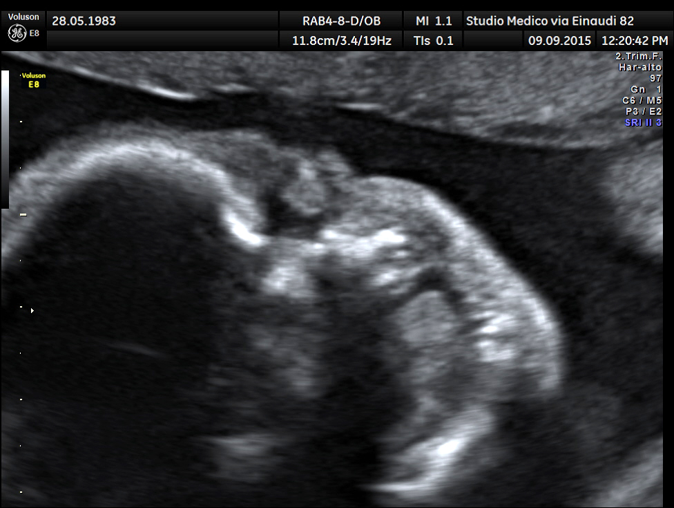
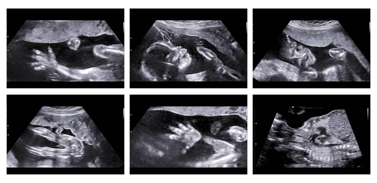
256 versus 128 Gray Scale Handheld Ultrasound Image Quality
An ultrasound image is built from shades of grey, since the machine turns the strength of each returning echo into a brightness, and the number of distinct grey levels it can show, two hundred and fifty-six or a hundred and twenty-eight, sets how finely it can separate one tissue from another by tone alone. A grayscale of two hundred and fifty-six means the image can carry that many steps from black to white, while a hundred and twenty-eight carries half as many, and the brochure presents the larger figure as the sharper, richer picture. It can be, where the difference shows, and it is also one of the more misunderstood numbers in the specification, because the eye, the display, and the noise in the image all conspire to decide how much of that tonal range a clinician ever truly sees. The grey-level count is a real quantity that matters in a narrow way, and reading it well means knowing exactly where it helps and where it is swamped by everything else.
More shades of grey can mean a richer picture, or a bigger number the eye never gets to use.
What grey levels do
To weigh the figure it helps to picture what the grey levels are for, since ultrasound carries its information almost entirely in tone, the brightness of each speck of tissue against its neighbours.
A structure shows up in ultrasound not by colour but by how strongly it reflects sound, so a slightly denser tissue returns a slightly brighter echo, and the image distinguishes it from the tissue beside it only if the two tones are different enough to tell apart. The grey-level count sets how finely those tones can be divided, so an image with more levels can place two faintly different echoes on two distinct shades where an image with fewer levels has to round them to the same one, blurring the boundary the clinician was looking for. Where tissues differ only subtly in echogenicity, a fine grey scale can reveal a border that a coarse one merges, and this is the honest case for more levels, the ability to separate things that lie close together in tone. Too few levels can also produce banding, where a smooth gradient of tissue is rendered as visible steps rather than a continuous shade, an artefact that distracts and can mislead, since a step that is an artefact of the grey scale can be read as a real edge in the tissue. The grey-level count, then, is at heart about contrast resolution, the ability to tell apart tissues that differ only a little in brightness, which is exactly the discrimination much of diagnosis rests on.
Ultrasound speaks in tone, and the grey levels are the size of its vocabulary.
Why the eye and the display cap the benefit
The complication that makes the grey-level number slippery is that the stored shades have to survive a long journey to a human eye, and several stages along the way discard the bulk of them.
The human eye, under ordinary lighting, can distinguish only a few dozen shades of grey at a glance, far fewer than even a hundred and twenty-eight, so the bulk of a fine grey scale is invisible to direct viewing and shows itself only when the operator adjusts the contrast to spread a narrow band of tones across the visible range. The display matters as much, since a phone or tablet screen in a bright room renders far fewer usable greys than its specification claims, and a grey scale finer than the screen can show is finer than the clinician can see. Ambient light makes this worse, washing out the darkest and lightest tones so the working range shrinks further, and the same image looks richer in a dim reading room than at a sunlit bedside. The value of a fine grey scale lies not in being seen all at once but in giving the operator headroom to adjust, since a stored image with many levels can be windowed and stretched to bring out a subtle difference, while one with few levels has nothing left to reveal when the contrast is pushed. The extra shades are a reserve the clinician draws on through the controls rather than a richness the eye perceives directly, and a maker that quotes the grey-level count as though it were visible detail has described the reserve as if it were the picture.
The eye sees a few dozen greys; the rest of the scale is headroom the controls can reach rather than detail the glance can catch.
The numbers that matter more than the count
Because the eye and the display cap how many greys are seen, the grey-level count is rarely the thing that decides whether an image looks good, and several other factors matter more.
The dynamic range of the captured signal sets how wide a span of echo strengths the image represents at all, and a probe that captures a narrow range and spreads it over many grey levels has fine steps across a small range, while one that captures a wide range gives the clinician more real information to work with regardless of the level count. Noise is the larger enemy, since speckle and electronic noise add a grainy mottle that obscures exactly the subtle tonal differences a fine grey scale was meant to reveal, so a noisy image with two hundred and fifty-six levels can show less true contrast than a clean one with fewer. The processing that turns raw echoes into the displayed picture, the filtering, the compression of the dynamic range, and the speckle reduction, shapes the visible contrast far more than the bare level count, and that is why two probes with the same grey scale can look entirely different. The grey-level count is a ceiling on contrast resolution rather than a guarantee of it, since the image can only use the levels the signal, the noise, and the processing leave meaningful. A buyer who fixes on the grey scale while ignoring the dynamic range, the noise, and the processing has read the easiest number and missed the ones that decide the picture, since the grey scale is the figure simplest to print and the others are the ones that take engineering to earn.
What it means for a handheld
A wireless handheld views its image on a phone or tablet in whatever lighting the work happens in, which makes the gap between stored grey levels and visible ones wider than it ever was on a cart with a calibrated monitor in a controlled room.
The handheld is used at the bedside, in the field, and in bright clinics where the screen is a consumer display under uncontrolled light, so the working grey range is squeezed by exactly the conditions the form was built for, and a fine stored grey scale buys less of its theoretical benefit than it would on a darkened reading station. This does not make the grey-level count useless, since the headroom to adjust contrast still helps the clinician pull a subtle difference out of a difficult image, but it does mean the count matters less than the quality of the processing and the noise control that decide how the image survives a bright screen. A maker that has invested in clean signal and good processing delivers a picture that reads well on a phone in daylight, while one that quotes a high grey scale over a noisy signal has printed a number that the viewing conditions erase. The buyer comparing handhelds should weigh how the image looks on the actual screen in the actual light far above the grey-level figure, since that is where the handheld’s picture is judged. The grey scale that survives a sunlit bedside counts for more than the one that only shines in a dark room.
Where the extra levels earn their place
It is fair to ask whether the finer grey scale ever changes a diagnosis, since a number that is mostly headroom can still matter at the edges, and the honest answer is that it helps in a few specific places and idles in the rest.
The work where contrast resolution is decisive is the work of telling apart tissues that differ only faintly in echogenicity, the subtle lesion against a similar background, the early change that has barely altered how a region reflects sound, and here the headroom of a fine grey scale lets the clinician window the image to bring the difference out. In high-contrast work, where the structures of interest are plainly brighter or darker than their surroundings, the extra levels do little, since the boundary is obvious at any reasonable grey scale and a coarse one separates the tones as well as a fine one. The benefit is real but narrow, concentrated in the difficult, low-contrast cases and absent from the easy ones, so a clinic that does mostly clear-cut work feels the grey scale less than one that hunts subtle tissue changes. Even where it helps, the benefit is conditional on the signal being clean enough that the subtle difference is not buried in noise, so the fine grey scale and good noise control have to arrive together to be of any use. A buyer who knows which kind of work they do can judge whether the grey scale is a feature that earns its place or a number that idles on their particular images, and a maker that asks what the buyer looks for is more useful than one that simply quotes the larger figure. The extra levels matter exactly where the diagnosis is hard and barely at all where it is easy.
The fine grey scale earns its keep on the subtle case and sits idle on the obvious one.
What the buyer should weigh
The grey-level count belongs in the comparison as a minor figure, and a buyer who keeps it in proportion judges the image better than one who lets it lead.
The first move is to look at the actual image rather than the number, ideally on the screen and in the light the probe will work in, since the picture is the only honest test of everything the specification tries to summarize. The second is to ask about the dynamic range and the noise performance, since those decide how much true contrast the image carries before the grey levels even come into play. The third is to keep the grey-level count in proportion, treating it as a ceiling that a good signal can use and a poor one cannot, rather than as a measure of quality on its own. The fourth is to judge the processing by eye, since the filtering and speckle reduction shape the visible contrast more than any single stored number, and only looking reveals whether they help or smear. A buyer who looks at the picture, asks about the signal beneath it, and keeps the grey scale in its modest place has read the image the way a clinician will, while one who chooses on the grey-level count alone has trusted the easiest number to print. Look at the picture, not the figure, and the grey scale falls into the small place it deserves, since the eye that will read the probe in service is the same eye that should judge it at purchase.
The image is the only number that matters in the end, and the grey scale is a footnote the eye quietly overrules.