





































Ultra-high frequency
18 to 24 MHz Ultra High Frequency Linear Ultrasound Probe Applications
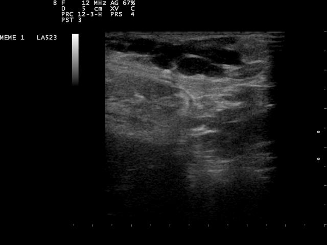
An ultra-high-frequency probe gives up more depth for detail than any other linear probe. Running at 18 to 24 megahertz, some higher still, it resolves structures finer than a tenth of a millimeter, reaching only a centimeter or two down. That suits whatever sits right at the surface, where the smallest differences decide the reading: the skin, the tiniest nerves and vessels, the eye. Where a standard linear probe sees a structure, this one reads its internal grain.
What the ultra-high band trades for
The physics that governs every probe runs hardest at this band. A frequency of 20 megahertz carries a wavelength under a tenth of a millimetre, so two structures that close together read as two rather than blurring into one. The detail is the finest ultrasound reaches. The same short wavelength is absorbed so fast that the beam fades within a centimetre or two of the surface. The field is tiny, and inside it the picture is the sharpest a sound beam draws.
That narrow window of detail and depth decides the whole use of the band. There is no point aiming an ultra-high-frequency probe at anything more than a centimetre or two down, since the beam never reaches it. The skill is in knowing which structures sit shallow enough to reward the resolution, and pointing the probe only there. The band is a specialist, built for one narrow job.
Reading the skin layer by layer
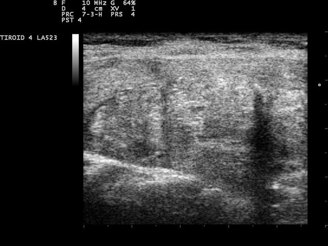
Dermatology is where the ultra-high band does its signature work. The skin is a layered organ, the epidermis over the dermis over the fat beneath, each a fraction of a millimetre to a few millimetres thick, and only this band resolves those layers as separate lines. A skin tumour reads against them, its depth into the dermis measured before it is cut, the figure a surgeon plans the margin from. A melanoma’s thickness, the single number that drives its staging and its treatment, can be estimated on the scan before the knife confirms it, so the operation is planned around a depth the probe read rather than a guess. An inflammatory skin disease shows its swelling and its altered layers, a scar shows its depth and how far it tethers the tissue under it, a cyst or a small lump shows its borders and what fills it. The probe maps the small vessels feeding a lesion, the flow that marks an active growth from a quiet one. On an ordinary probe the whole skin collapses into one bright band a millimetre or two thick. The ultra-high band opens that band into its parts, the layers a knife will cross laid out before the first incision. The depth it reaches is small. The skin lives entirely inside that depth, so the band reaches the whole of the organ it is pointed at. At the surface itself, that shallow reach is all the depth the work needs. The depth the probe reads is itself the clinical number. A tumour two millimetres into the dermis and one four millimetres deep call for different operations, and the band measures that difference where the eye on the surface cannot. The figure off the scan sets the margin and the depth of the cut before the skin is ever broken, which is why the band has become a planning tool and not only a diagnostic one.
The smallest nerves and vessels
The band reaches nerves and vessels too fine for an ordinary probe. A digital nerve in a finger, a millimetre across, shows its course and where it is cut or trapped, a read that guides a repair. A small vessel feeding a flap or a fingertip shows its flow, the patency a reconstructive surgeon checks before and after an operation. A tiny foreign body, a glass splinter a fraction of a millimetre wide lodged just under the skin, throws a reflection the band catches and brings out clearly on the picture.
A peripheral nerve reads here as its individual fascicles, the fine internal structure that shows whether a nerve is healing cleanly or scarring shut. The resolution turns a nerve from a single grey cord into a structure a surgeon reads the inside of. That detail matters in the hand and the wrist, where the nerves are small and the stakes of a repair are high.
The band reads the microvasculature too. The fine vessels in a skin flap or a fingertip show their flow on power Doppler, the perfusion a reconstructive team watches to know a flap has taken. A vessel a fraction of a millimetre wide carries a faint signal the band is sensitive enough to draw.
The newest machines push this further with microvascular imaging, a mode that strips the background clutter to map the finest flow, vessel by vessel. On the ultra-high band that mode reads the perfusion of a structure a millimetre across, the tiny vessels inside a tumour or a healing flap drawn vessel by vessel. The density of that flow grades a lesion or confirms a graft has taken, a count the surgeon acts on.
The eye and the front of the body
The eye sits in the band’s range and rewards it. The cornea, a fraction of a millimetre of layered tissue, reads on the highest-frequency probes, its thickness measured and its layers seen. The front chamber of the eye, the iris, the lens surface, each sits shallow enough for the band to draw. An ophthalmic ultra-high probe reads structures a general probe leaves unresolved.
The band is the tool of ultrasound biomicroscopy, the high-frequency scan of the eye’s front. It reads the angle where the iris meets the cornea, the depth of the anterior chamber, a cyst or a tumour on the iris, the structures that sit behind what a slit lamp can show. The scan plans an implant and follows a glaucoma angle, a read of the eye’s drainage that the surface examination alone leaves hidden.
The surface of a joint, the lining of a tendon sheath, the smallest lymph nodes, each sits in the shallow field the band owns. A rheumatology service reads the fine erosions at a joint surface and the thin inflamed lining that marks early disease, changes a millimetre or less across, the fine grain the band is built to hold. The band reads the body’s outermost layer wherever fine detail there changes a decision.
The nail unit reads on the band too, the matrix and the bed under the plate. A small tumour or an inflammation there shows on the band, the depth measured before a procedure. A glomus tumour under the nail, a few millimetres across and exquisitely tender, reads as the small dark mass it is, marked for the surgeon before the nail is ever lifted.
A temporal artery, thin and right under the skin, shows its wall on the band. The thickening of an arteritis reads on the scan, pointing a biopsy to the segment worth taking, the imaging standing in for a blind cut.
How the band reaches its detail
The detail comes from the wavelength and the way the probe is built. At 20 megahertz the wavelength in tissue runs about 75 microns, so two structures that far apart still read as two, near the limit of what sound resolves. The crystal is cut thin to ring this high and its elements are packed close, and some ultra-high probes use a different transducer entirely, a capacitive micromachined element that rings cleanly across the wide band. The engineering is what lets the probe reach a frequency a standard linear face cannot.
The shallow field comes with its own gains. A picture only a centimetre or two deep is swept fast, the frame rate high enough to follow a vessel pulsing or a tendon gliding in real time. The short pulse that draws the fine grain also gives a clean line along the beam, the detail in depth as fine as the detail across it. The reach is the cost, the high frequency absorbed within a centimetre or two, so the probe is pressed gently with plenty of gel to couple its sound cleanly into the skin.
What the resolution costs nothing for
For work at the surface, the band’s shallow reach is all the depth the job asks for.
Where the ultra-high probe fits a kit
Several services carry this band, each for work that lives in the first centimetre or two. A dermatology service uses it for lesion depth and margin planning, the read that shapes an operation. A reconstructive or hand surgeon turns to it for the small nerves and vessels a repair hangs on, an ophthalmology clinic for the cornea and the front of the eye, a rheumatology service for the fine joint-surface changes of early disease. For each, the band’s resolution reads what no other probe resolves.
The probe itself is built small to match the work. A compact footprint, sometimes a hockey-stick shape with the face set at an angle, reaches a fingertip, a nail fold, the corner of an eye, the tight spots the surface work lives in.
The image setup leans on the band’s strengths. A shallow depth fills the screen with the first centimetre, the line density draws the fine grain, and the focus sits near the surface where the structure is. The machine is set for the top layer, not for the depth a standard scan reaches toward.
The band asks something of the operator in return. The structure is so shallow that a heavy hand flattens it, so the probe is floated on a bed of gel with the lightest touch, a standoff pad sometimes laid between probe and skin to lift a surface structure into the focal zone. The narrow window of depth leaves no room for a loose setup, since a band this fine shows every error in the focus or the gain as plainly as it shows the anatomy.
The band rarely travels alone. It joins a standard linear probe in a kit, the ordinary band for the work a centimetre or more down and the ultra-high band for the surface detail above it. A clinician reaches for the ultra-high probe when the question turns on the finest grain at the surface itself, and for the standard one when the structure sits deeper. The two cover the superficial field between them, the ultra-high band holding the top layer that the standard band reads through.
The handheld form brings that resolution to the bedside. A pocket ultra-high-frequency probe reads a skin lesion in a clinic room, a digital nerve in a hand unit, the cornea at a bedside, the picture a cart-bound machine once held drawn in one hand against the surface of the body.
The band has a place among the other ways to read the surface. A dermatoscope reads a lesion’s surface in light and sees nothing of its depth, and optical coherence tomography reads the top fraction of a millimetre in fine detail before it stops. The ultra-high band reaches through the whole thickness of a lesion and the tissue beneath it, the depth those light-based tools stop short of, on a machine already in the clinic for other work. It fills the gap between what the eye and the light tools catch at the surface and what a standard probe reads in the layers below, the three together reading the skin from its surface down to its base.
Common questions about ultra-high-frequency ultrasound
What frequency is ultra-high-frequency ultrasound?
Roughly 18 to 24 megahertz, with some probes reaching higher. It sits a step above the ordinary high-frequency band and pushes the resolution finer than a tenth of a millimetre.
How deep does an 18-to-24 MHz probe see?
Only a centimetre or two. The short wavelength that gives the fine detail is absorbed within that depth, so the band is built for structures right against the surface, not for anything deeper.
What is it used for in dermatology?
Reading the skin layer by layer: a tumour’s depth into the dermis for margin planning, a melanoma’s thickness before surgery, an inflammatory disease’s swelling, a scar’s depth, and the small vessels feeding a lesion.
Why does it suit small nerves and vessels?
It resolves a structure a millimetre or less across, so a digital nerve shows its course and fascicles, a small flap vessel shows its flow, and a tiny foreign body throws a reflection a coarser probe would miss.
Can it image the eye?
Yes. The cornea, the front chamber, the iris, and the lens surface sit in its shallow range, and the band resolves the corneal layers and thickness a general probe cannot.
Does it replace a standard linear probe?
No. It joins one in a kit. The ultra-high band reads the top layer at the surface; the standard band reads the structures a centimetre or more down. The two cover the superficial field between them.

About the Author
Julien Mercier
Senior R&D Engineer · Medical Ultrasound Transducer Development
Senior R&D Engineer with an M.S. in Applied Physics and over 15 years of experience in medical ultrasound transducer development, specializing in the design verification and performance testing of high-frequency imaging transducers. Currently leading the development and verification of the company’s next-generation high-frequency linear-array transducer, responsible for imaging performance evaluation and reliability analysis in preclinical testing. Brings extensive hands-on experience in piezoelectric element tuning, beamforming parameter optimization, and system-level performance testing.