Superficial Ultrasound Linear Probe Wireless Imaging
Superficial ultrasound reads the body in the first few centimetres under the skin, where a high-frequency linear probe draws the sharpest picture an ultrasound machine can make. Everything from a thyroid to a tendon sits within that reach, rendered in fine detail, with a wash of color over the picture showing the blood that moves through it. This is the foundation every superficial exam is built on. The frequency, the probe, and the Doppler mode are the few choices that decide what a scan can see.
What the first centimetre holds
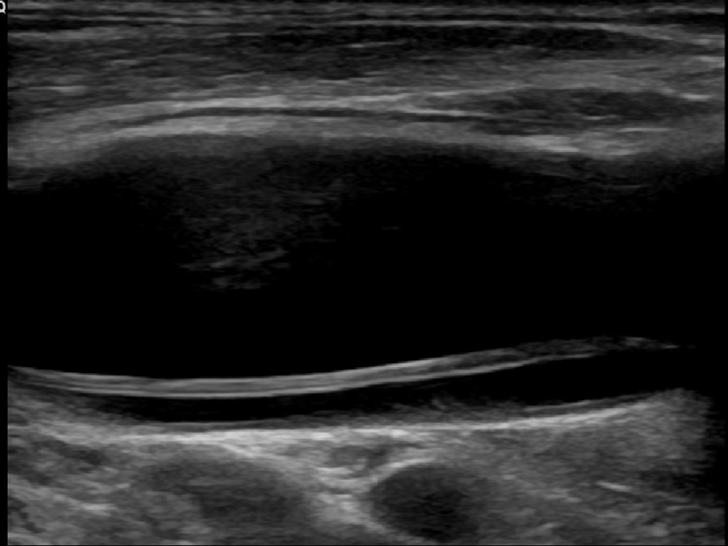
Two vessels of the neck, seen as dark channels under the skin with a high-frequency linear probe. A vessel reads as a black tube against the gray tissue, the look that lets a reader trace it, press it to tell a vein from an artery, or watch a needle cross into it. Image: Mme Mim, CC BY-SA 4.0.
High frequency is what makes the surface picture sharp. Sound at twelve or fifteen megahertz draws a far finer grain than the low pitches a deep scan needs, fine enough to split the layers of a tendon or the wall of a small vein. Resolution comes in two directions, along the beam and across it, and a high frequency sharpens both: shorter pulses down the line of sound, a tighter beam to either side. A fifteen-megahertz linear probe resolves detail well under a tenth of a millimetre, the scale of a vessel wall or a single tendon fiber. The detail holds steady enough to follow a change over time, the same nodule or node measured the same way on a later visit and read against the first. A handheld measures all of this on the screen, the calipers placed with a fingertip and the number frozen on the image. The price of that frequency is reach. Sound loses strength along its path, and faster the higher the pitch, roughly a decibel for each centimetre and each megahertz. A seven-megahertz wave still carries a few centimetres into the neck. Climb to twenty and the same sound is gone within one. The linear probe holds to what lies near the skin and leaves the deep body to other heads.
What a high-frequency probe brings into view in that first centimetre is a world of small structures. A tendon shows as a tight grain of bright parallel fibers. A vessel shows as a dark channel that fills with color when the flow is switched on. A gland shows as an even, speckled gray. A nerve shows as a cluster of bright dots in a dark sheath. A reader learns these as the look of healthy tissue, so a lump, a tear, or a swelling stands out against them at a glance. Each tissue carries its own texture on the screen. Muscle reads as a feathered weave. Fat reads as a soft, layered gray. A lymph node reads as a dark bean with a bright fatty centre. The trained eye reads the whole picture as a pattern, and notices the break in it before any measurement is made.
The detail is fine enough to measure on. A nodule is sized to the millimetre, a tendon followed fiber by fiber, a needle watched as a bright dot crossing into a vein. None of this is possible at the lower frequencies a deep scan runs, where the same structures blur into a soft gray. The surface is where ultrasound draws its cleanest line. The range it covers is wide. The thyroid and its nodules, the breast and its lumps, the lymph nodes of the neck and groin, the muscles and tendons of a sore joint, the nerves a block must find, the vessels a line must enter: each sits within the linear probe’s reach, and one probe with one set of habits carries across the whole of it. Much of it reads best in motion. A tendon glides under the skin when the joint bends, a muscle thickens when it works, a vessel pulses with the heart. The probe holds the picture live, so a tear that hides at rest opens under load and a flow too faint to see steadies under a held breath. The shallowest targets sit so close to the face that they need a little distance to come into focus, and a pad of gel or a built-in standoff lifts the skin a few millimetres into the beam’s sharp zone.
How high the frequency runs
The frequency is the first dial a reader sets, and it sets the whole character of the scan. A general superficial probe runs in the band the field uses for the bulk of surface work, high enough to read the thyroid and the vessels in sharp detail, with enough reach left to hold a structure a few centimetres down. The choice within that band tunes the scan toward depth or toward detail, a small shift a reader makes to suit the organ in front of them. The standard band runs roughly from seven to fifteen megahertz, and the machine slides within it from a preset, lower for a deeper thyroid or a larger patient, higher for the finest grain on a shallow target. A reader rarely thinks in numbers, picking the organ and letting the software load the rest. Depth is read off a scale down the side of the screen, and a superficial study rarely runs it past four or five centimetres, the picture staying sharp across that window. The focus marker sits at the depth of the target, where the beam pulls tightest and softens above and below. The one linear head stretches across its whole band in software, so a single probe reads both the shallowest gland and a muscle a few centimetres down. Modern probes squeeze more out of a crystal than the raw frequency suggests. Harmonic imaging listens at twice the sent pitch, clearing haze and sharpening an edge with no new probe. Compound imaging sweeps the beam at several angles and averages them, smoothing the speckle a single angle leaves. The number printed on a probe is its centre frequency, the middle of the band it works across, and a head labeled twelve megahertz runs usefully from below ten to above fifteen.
For the shallowest work there is a higher band again. Ultra-high-frequency probes, running past eighteen megahertz, read the skin’s own layers and the fine vessels of the face, a depth of a centimetre or less drawn in a detail the standard band cannot reach. This is the probe of dermatology and aesthetic medicine, where the target sits a hair under the surface and a millimetre of depth is a long way down. The reach shrinks at the higher frequencies, so the ultra-high head trades the body’s depths away for a picture of the skin no other probe can match. In practice a clinic owns one or two linear heads and runs them across the whole superficial list. A single broad-band linear probe, tuned by software, covers the thyroid, the breast, the vessels, and the musculoskeletal work from one body. The ultra-high head is a second buy, reached for only when the target sits in the outermost millimetre of skin.
The flat face
A linear probe is flat across its face. It fires its lines of sound straight down side by side and returns a square picture, the same width at the top and the bottom, with no fan to it. That square field suits a structure lying flat under the skin, read across its whole length in one view. A long tendon, a stretch of vessel, a row of lymph nodes: each is laid out end to end on the screen at a size the eye can read. Behind the flat face sits a row of small crystal elements, a hundred and twenty-eight of them or more, fired in groups to build the picture one line at a time. The number of elements sets how fine and how steady the picture can be drawn, one of the quiet specifications that separates a probe a clinic trusts from one it works around. In front of the elements sit a matching layer and a lens. The matching layer eases the sound out of the hard crystal and into the soft skin, so less of it bounces back at the face. The lens focuses the beam to a thin sheet at the working depth. Both are fixed in the probe, the reason a head suits one band and one depth and reads best within them.
How the flat face stands against a curved one comes down to the depth a clinic needs. The flat linear face reads the surface in fine detail. A broad curved face reaches the deep belly through a wide fan. A small curved face slips a sector beam into a tight space between bones. A clinic that scans both the surface and the depths carries more than one head on the same base, the linear for the skin among them. The flat face has its own limit. It needs a flat window of skin to sit against, and it struggles over a curved or bony surface where its edges lift off and lose contact. A small joint, a finger, the hollow behind an ankle: these sometimes ask for a smaller head or a generous pad of gel to keep the whole face in touch. The square picture earns its place where it sits flat: a structure keeps its true shape and size clear across the width of the image, with none of the stretch a fan-shaped field adds at its edges. The width of the face sets how much of a structure fits in one view, a wider head taking in a whole thyroid lobe where a narrow one shows it in parts.
Color over the picture
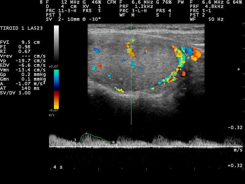
A thyroid on a 12 MHz linear probe with color Doppler laid over it, the orange and blue marking blood that flows toward and away. The trace along the bottom is the pulsed-wave waveform from the gated spot. The figures down the side are its velocity and resistance readouts. Image: Nevit Dilmen, CC BY-SA 3.0.
A wash of color Doppler over the gray picture marks where blood flows and which way it runs, turning a still anatomy picture into a reading of flow. A vessel fills with color where blood moves through it. A solid lump shows the vessels that feed it. A clot shows as a gap where the color should be, the flow filling the open channel and stopping dead at the blockage. This is the second half of the superficial scan, the half that reads the blood moving inside the tissue. Doppler reads flow by the shift in the echo’s pitch. The size of the shift gives the speed of the blood, the sign of it gives the direction, and the machine paints both as color over the gray. The reading leans on the angle. Flow straight across the beam returns no shift at all, so a reader tilts the probe to bring a vessel more along the line of sound before trusting the number.
Choosing between color and power Doppler turns on the question in front of the reader: the direction a vessel runs, or the first faint trace of flow in an inflamed gland. Color codes the flow toward and away in two colors. Power paints any flow at all, blind to direction, sensitive down to the slow trickle a color map would miss. A reader switches between them in a tap. Color asks for its own settings. The box of color is kept small and laid right over the vessel, since a wide box slows the picture. The scale is matched to the speed of the flow, so the color fills cleanly with no smearing. Color is read on top of a good gray picture, never in place of one. A reader sets the gray image first, finds the vessel or the lump cleanly, and only then lays the color over it. Power Doppler earns its place on the inflamed and the small: the flushed rim of an infected joint, the busy core of an active nodule, the faint feed of a tiny lump, each lit up where a color map shows little. Doppler in every mode needs the flow running along the beam to read its speed, and a vessel crossed square returns no shift at all.
A third mode reads a single vessel as a trace. Pulsed-wave Doppler drops a gate onto one chosen point and draws the speed of the blood there as a waveform, a hard number for a flow the color wash only paints. The height of the wave gives the peak speed. The shape of it tells a fast jet from a slow drift, the rhythm of the pulse, the resistance downstream. It is the mode that turns a color picture into a measurement.
The effects the picture throws
Ultrasound draws a few effects of its own, and a reader learns to expect them. A stone or a patch of bone throws a dark shadow behind it, where the sound cannot pass through. A fluid-filled cyst brightens the tissue behind it, since the sound crosses the clear fluid losing nothing. Neither is a fault in the picture. Read rightly, each is a clue to what sits in the image, a shadow behind the hard, a bright tail behind the fluid.
A needle shows a signature of its own. The metal returns a bright dot or line where it sits, with a ladder of fainter copies stacked below it, the echo ringing back and forth inside the steel. A reader reads the top bright mark as the tip and the ladder as its echo, the habit that keeps a needle in view on its way into a vein. A pocket of air does much the same, throwing a bright streak and a dirty shadow that a reader learns to tell from solid tissue.
Each effect has a name and a cause, and a reader meets them early, since the surface throws them constantly. A scan misread for its artifacts finds disease where there is none, or misses it where it hides behind a shadow. A reader reads past an artifact to the tissue under it, dropping the probe to a new angle to clear a shadow or to confirm a dark patch. Knowing the shadow, the bright tail, the anisotropic dark patch, and the needle ladder is part of reading the surface at all. These are the picture’s own habits, and a reader who knows them is rarely fooled by one. What separates a clean superficial scan from a poor one is rarely the machine. A probe held square, a gain set even, a focus dropped to the target, a structure read in two planes: these settle the picture more than the cost of the hardware does. A modest probe in a careful hand reads the surface well.
Reading near the surface
The method behind every superficial scan is the same handful of moves. Set the depth shallow until the target fills the screen, drop the focus marker to its level, lay the probe flat with a film of gel, and sweep through the structure in two directions at right angles. A lump is measured in three planes. A vessel is followed along its length and across it. The gain is set so the tissue reads in an even gray, neither washed white nor sunk dark. The focus marker is dropped to the depth of the target, where the beam pulls tightest. Pressure is a tool of its own: a gentle press flattens a normal vein, the simplest test of whether it is open or clotted, run all the way down a swollen leg to hunt the spot it will not give.
One habit matters more than any setting. Sound returns brightest when it strikes a structure square on, so a tendon or a nerve can darken and seem to vanish when the probe tilts a few degrees off. A reader learns to rock the probe until the target lights up, and to trust a dark patch as real only once it holds dark through that small sweep. This one trick separates a clean superficial scan from a confusing one.
The same settings carry from a thyroid in the morning to a tendon in the afternoon, the depth and the focus nudged a little for each. This shared method is why a single linear probe, in a trained hand, reads the whole range of surface organs from the same few habits. The gain is set for an even gray, neither washed white nor sunk dark, with a light hand on the probe and a look from more than one angle before a finding is called. Orientation is a discipline of its own. The probe marker stays to a fixed side so the screen matches the body the same way every time, the left of the image the same side of the patient on every scan. Every superficial study ends in a record: a structure measured in its planes, the numbers frozen on the image, the clip labeled with the side and the site and saved to the patient’s file in a tap. A reader builds speed with the routine, a full superficial sweep settling into a few minutes once the sequence is second nature. The same flat probe guides a needle as readily as it reads tissue. A vein is held in the centre of the screen, the needle laid in the plane of the beam, its bright tip watched all the way into the vessel. The surface is where guidance is easiest, the target shallow and the needle in clear view, the reason a linear probe is the one a clinician reaches for to place a line, drain a collection, or sample a node.
On a wireless handheld
The wireless form puts this whole range into a pocket. A linear head the size of a thick pen runs from a phone, the processing inside the probe and a radio link in place of a cable. The work that once filled a cart now sits in the probe’s handle. A chip forms the beam, builds the image, and runs the Doppler, then sends a finished picture to the phone over the link. The phone adds the screen, the storage, and the way out to a colleague or a patient record. The same phone stores the scan, measures on the screen, and sends a thyroid or a breast image to a specialist for a second read in seconds. A clinic far from an imaging department reads a vein, a lump, or a tendon in the room where the patient sits, on a device that costs a fraction of a cart. The whole of superficial ultrasound, built on these few fundamentals, now travels wherever a patient lies. A wireless probe is a complete machine, not an accessory. The crystal, the electronics, the battery, and the radio live in one sealed body the size of a marker, and the phone behind it carries no ultrasound hardware at all, only the screen and the software. The battery runs a session of an hour or two and charges between patients. Heat builds in the sealed body through a long scan, and the probe eases off before it warms in the hand. The link reaches past the room: a clip saved on the phone travels to a specialist across the city for a second read, or guides a junior’s hand by voice from a senior on a call. The price has fallen with the size, putting a sharp superficial picture within reach of a single doctor or a small clinic for the first time. A clinic far from any imaging department reads its own surface scans on the spot, an answer given in the room where the patient sits. The whole device recharges on a small cradle between clinics, ready for the next list by morning.
Common questions about superficial linear-probe ultrasound
What is a linear ultrasound probe used for?
Reading the structures near the skin. A flat, high-frequency linear probe shows the thyroid, the breast, the vessels, the nerves, the muscles and tendons, and the small parts in the first few centimetres under the skin. It draws them in fine detail and lays color over them to show the blood flowing through.
What frequency does a superficial scan use?
High, somewhere above seven megahertz. A general superficial probe runs in the band the field uses for the thyroid, the breast, and the vessels. The shallowest work, the skin’s own layers, calls for an ultra-high band past eighteen megahertz. A higher frequency draws finer detail and reaches less far into the body.
What is the difference between color and power Doppler?
Both paint blood flow in color over the gray picture. Color Doppler codes the direction the blood runs, toward or away. Power Doppler ignores direction and catches the faintest flow of all, down to the trickle of an inflamed gland or a tiny vessel. A reader reaches for color to read direction, for power to read presence.
What is pulsed-wave Doppler?
A mode that reads the speed of blood at one chosen spot. It drops a gate onto a single point in a vessel and draws the speed there as a rising and falling waveform. The shape and the height of that wave tell a fast jet from a slow drift, and put a hard number on a flow the color wash only shows in color.
Can a handheld linear probe replace a hospital ultrasound machine?
For the focused superficial question, it comes close. A good linear handheld reads a thyroid nodule, a breast lump, a vein for a line, or a torn tendon to a standard a cart once held. The finest studies and the deep-body scans stay with a full system and a trained sonographer.
About the Author
Julien Mercier
Senior R&D Engineer · Medical Ultrasound Transducer Development
Senior R&D Engineer with an M.S. in Applied Physics and over 15 years of experience in medical ultrasound transducer development, specializing in the design verification and performance testing of high-frequency imaging transducers. Currently leading the development and verification of the company’s next-generation high-frequency linear-array transducer, responsible for imaging performance evaluation and reliability analysis in preclinical testing. Brings extensive hands-on experience in piezoelectric element tuning, beamforming parameter optimization, and system-level performance testing.