Superficial Linear Color Doppler Wireless Ultrasound Probes
A linear probe reads the body in the first few centimetres under the skin and draws it sharper than any other head can. That shallow band holds a surprising amount of medicine, from the thyroid in the neck to a vein before a needle. A wash of color over the gray picture shows the blood moving through any of it. On a wireless handheld, that whole reach travels to the bedside, the clinic, and the rural outpost.
The linear probe and its shallow reach
The linear probe reads the surface in fine detail. Its face is flat, so the lines of sound run straight down side by side and the picture comes back as a square block. Its frequency runs high, somewhere from seven to fifteen megahertz and higher on the finest probes. High-frequency sound draws the sharpest picture the machine can make, fine enough to separate the layers of a tendon or the wall of a small vein. The price of that frequency is reach. The sound fades within the first few centimetres, so the linear probe holds to what lies near the skin.
That shallow range is exactly where a great deal of medicine lives. The thyroid sits a centimetre under the neck. A breast lump, a swollen lymph node, a vein before a needle, a nerve before a block, a torn tendon: each rests within the linear probe’s reach, close enough that the high frequency renders it in crisp detail. The probe is held flat against the skin with a film of gel. The structure underneath then lies across the screen at a size the eye can read. A small thing a hand can barely feel fills half the screen under a high-frequency head.
The image itself asks for a steady hand and a careful angle. Sound bounces back brightest when it strikes a structure square on, so a tendon or a nerve can darken and seem to vanish when the probe tilts a few degrees off. A reader learns to rock the probe until the target lights up, and to trust a structure only once it holds its brightness through that small sweep. This habit, more than any single setting, is what separates a clean superficial scan from a muddled one, the first thing a beginner on the probe builds.
Color over the gray picture
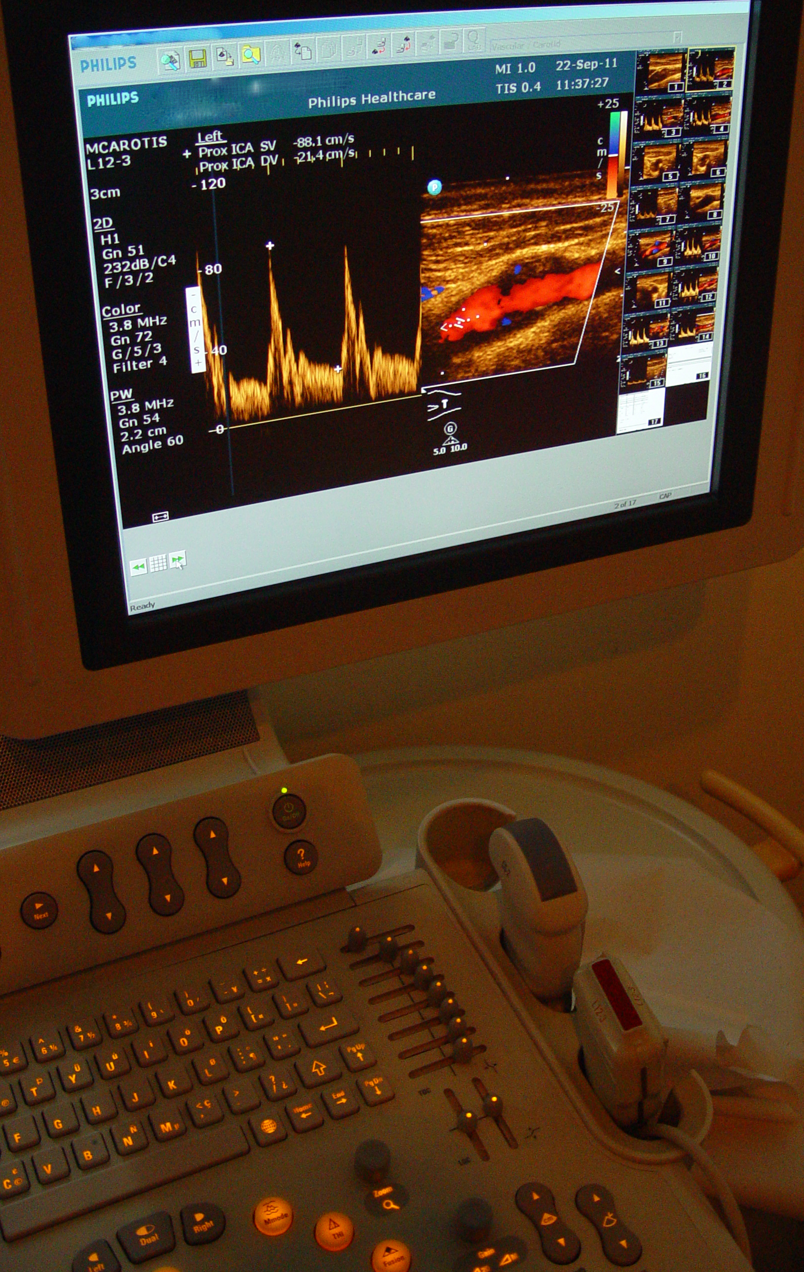
A carotid color-Doppler study on a conventional system. The red wash over the vessel is blood in flow; the yellow trace below is the spectral waveform, read for the peak speeds on the left. The probe on the console is a linear one, the “L12-3”; the “MI” and “TI” figures are the machine’s safety readouts. A wireless handheld carries the same color and trace in a pocket. Image: Etan J. Tal, CC BY 3.0.
The gray picture shows the shape of things. A wash of color Doppler laid over it paints the one thing the gray leaves out: the blood in a vessel, which way it runs, and how fast. This is the half of the scan the word color names, the half that turns a still anatomy picture into a reading of flow. A vessel fills with color where blood moves through it. A solid lump shows the vessels that feed it. A clot shows as a gap where the color should be and is not. The same wash tells a living vessel from a scarred one and a feeding artery from a draining vein.
Power Doppler is the more sensitive setting. It drops the sense of direction and paints any flow at all, down to the slow trickle in a small vessel or an inflamed gland. A thyroid nodule lit up with power Doppler, a tendon sheath glowing with it, a lymph node flushed through the middle: each tells a reader that something is active in the tissue. Between the gray detail and the color flow, the linear probe answers two questions at once, what a structure is and whether blood is moving through it.
A third setting reads flow as a trace. Spectral Doppler drops a gate onto one vessel and draws the speed of the blood there as a rising and falling waveform. The height of that wave gives a peak speed. Its shape separates a vein from an artery, an open vessel from a narrowed one. The bulk of superficial work leans on the color wash. The trace waits for the moment a number is needed.
Color does real work across the whole superficial range. It tells a cyst with no flow from a solid lump fed by vessels. It flags the angry flush of an infected joint or an inflamed gland. It confirms a vein is open before a needle goes in, and finds the clot that fills a swollen limb. In the testis it carries the gravest reading of all, the flow that is there or gone in a few painful hours.
The everyday superficial scan
The bulk of the linear probe’s work is the same handful of moves repeated on a new structure. Set the depth shallow, drop the focus to the target, lay the probe flat, and sweep through the structure in two directions at right angles. A lump is measured in three planes. A vessel is followed along its length and across it. The general method, the depth and focus and the order of the sweep that every superficial scan is built on, carries from one organ to the next with only the target changed. The settings are few, the same on every scan. The depth is wound down until the target fills the screen, the focus marker dropped to its level, the gain set so the tissue reads in an even gray. A high-frequency preset loads the rest. Once these are set for the thyroid, the same handful carries to the breast or the vein with only a nudge, which lets one probe move through a clinic’s whole superficial list in a morning. A thyroid, two breasts, a swollen node, and a sore shoulder run on one head without a change of probe.
The thyroid in the neck
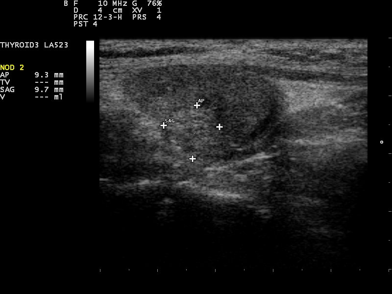
A thyroid nodule on a linear-probe scan, measured. The rounded darker patch in the gland is the nodule; the dotted crosses are the operator’s calipers, sizing it at 9.3 by 9.7 millimetres. The “LA523” and “10 MHz” note the linear probe and its high frequency. Image: Nevit Dilmen, CC BY-SA 3.0.
The thyroid is the linear probe’s home ground. It sits shallow, wraps around the windpipe, and shows up in fine detail under a high-frequency head. The everyday question is the nodule: a lump in the gland, common and usually harmless, read for the few features that mark the rare one needing a closer look. A reader weighs whether a thyroid nodule looks calm or suspicious by its shape, its edge, its echoes, and the flecks of calcium inside it, then measures the gland and counts the nodes in the neck beside it.
The read follows a checklist the field has settled on. Height greater than width, a ragged edge, a core darker than the gland around it, bright specks of calcium inside: each feature adds to a nodule’s score. The score sets whether it goes to a needle or to a yearly watch. The aim is narrow, to find the few nodules that earn a closer look and to leave the many that do not.
The gland itself tells its own stories. A thyroid gone patchy in its texture reads as the inflammation of thyroiditis. One studded with nodules and grown large is a goiter pressing on the neck. The flow through the gland reads high in an overactive thyroid, a clue the color picks up at a glance. A reader takes in the whole gland before settling on any single lump.
Color adds the last layer. A nodule may carry its own tangle of vessels. The pattern of that flow feeds into the read. The whole study runs in minutes at the bedside, the gland measured, the nodules sized and scored, the neck swept for a swollen node. A handheld with a good linear head does this work as well at a rural clinic as a cart does in a hospital room.
The breast
Breast ultrasound asks a sharper question: is a lump a simple cyst or something solid. A cyst is a smooth black pocket of fluid that the sound passes straight through. A solid lump holds its own gray and casts no clear space behind it. Telling the two apart is the first job. The linear probe does it in seconds, where a hand feels only a vague fullness. The full read of a lump found in the breast weighs its shape, its edge, its depth against its width, and the shadow or the flow around it.
The scan shifts to suit the breast it reads. In a young or a dense breast, where the tissue is dense, ultrasound comes into its own, reading lumps a mammogram tends to hide. In an older breast it works alongside the mammogram, sorting the lumps the X-ray flags. When a lump needs sampling, the probe holds it still for the needle that takes a core, the tip watched into the target. The same picture that finds the lump guides the test that names it.
The scan earns its place where mammography reaches its limit. Dense breast tissue hides a lump on a mammogram. On ultrasound that same lump can stand out plainly. The probe also guides a needle into a lump to sample it, the target and the needle held in one picture. For a clinic far from an imaging department, a handheld linear probe brings the first answer to a frightened patient in the room where she sits.
The small parts
A scatter of small organs near the surface falls to the same probe. The salivary glands in the cheek and under the jaw, swollen by a stone or an infection. The lymph nodes along the neck, the armpit, the groin, read for the shape that tells a reactive node from a worrying one. A lump in the fat under the skin, sorted into a cyst, a lipoma, or something that needs a closer look. A lymph node gone round, with its bright fatty centre lost, earns a second look. The testis is the urgent one among them: a sudden painful testis is scanned for the flow that decides whether it is twisted and dying or merely inflamed. Color reads both testes at one setting and holds them side by side, the surest way to catch a missing flow before the hours run out. These structures share a way of reading. Each is shallow, each is small, and each turns on the same questions of shape, edge, and flow. The full range of the small parts near the surface sits within a single probe’s reach, scanned with the same flat head and the same wash of color.
The needle and the vein
The linear probe changed how a line goes into a vein. A clinician once found a deep vein by feel and landmark and hoped the needle struck it. Now the probe holds the vein on the screen and the needle is watched the whole way in, the tip kept in view as it crosses the skin, the wall, and the lumen. A miss, a near artery, a vein too small or clotted: each is seen before the needle moves.
The method runs two ways. The probe sits across the vein, the needle entering from the side as a bright dot through the wall. Or it lies along the vein, the whole shaft followed down to the tip. Either way, placing a line under ultrasound turns a blind stick into a watched one, which is why it is now the standard for a central line and a hard peripheral one alike. A handheld puts that safety in any room a patient is cannulated in.
Before the needle, the probe sorts the vessels by a gentle press: a vein gives way and flattens, the sign that marks it from the artery beside it. The same press, run down the length of a swollen leg, hunts the spot a vein will not flatten, the mark of a clot lodged inside. Color confirms what the press suggests, the flow filling an open vein and stopping dead at the clot that fills the swollen leg.
Finding a nerve for a block
Regional anesthesia leans on the same picture. To numb an arm or a leg for surgery, the anesthetist lays the drug around a nerve. The linear probe is how the nerve is found and the needle steered to it. A nerve shows as a bundle of bright dots in a dark sheath, told from the vessels and tendons that lie near it by its look and the way it holds through a tilt. The needle is followed to the nerve’s edge. The drug is watched going in. The nerves a handheld reaches are the big named ones near the surface, the plexus above the collarbone for an arm, the femoral and the sciatic for a leg.
The spread is the proof. Anesthetic pushed in the right plane opens a dark pool that wraps the nerve in a ring, the sign that the block will take. A pool on one side only, or no pool at all, tells the operator to move the needle before more drug goes in. Working out where to place a nerve block and reading that spread is a skill the handheld brings to the edge of the operating room and the emergency department alike.
Muscle, tendon, and joint
The moving parts read well under a linear probe because so much of the joint lies shallow. A tendon shows a fine grain of parallel fibers when it is whole. A tear breaks that grain into a dark gap. A muscle shows its feathered pattern, a joint its lining and the fluid that gathers when it is inflamed. The probe reads these live, so a tendon is watched as it glides and a muscle as it contracts, a motion a still picture would miss, the reason so much of this work is read in real time.
The catch in reading a tendon is its trick of vanishing. A tendon throws back a bright signal only when the sound strikes it square, so a tilt of a few degrees turns it dark and mimics a tear. A reader learns to rock the probe and watch the tendon brighten, calling a dark patch a tear only once it stays dark through the sweep. This one artifact sets off more false alarms than any real disease, the first trap a reader is taught to avoid.
A torn tendon can look whole at rest, then pull apart when the muscle behind it works, so the probe watches it live as the joint moves. A joint swollen with fluid shows a dark pocket the probe can mark for a needle. A tendon slipping over a bone, a muscle tearing under strain, a nerve catching at a joint: each shows in motion what a frozen frame would hide.
Much of this is bedside work with a fast answer. A swollen joint is checked for fluid before a needle drains it. A painful shoulder is scanned for a tear in the tendon that a hand cannot feel. The same probe that reads the thyroid in the morning reads a torn calf in the afternoon, the depth and the focus shifted a little and the reading eye carried across.
Why wireless and handheld
The wireless form is what carries the whole superficial range into a pocket. A linear head the size of a chunky pen runs from a phone, with the processing in the probe and a radio link in place of a cable. The same phone stores the scan, measures on the screen, and sends a thyroid or a breast image to a specialist for a second opinion. A device this small reads a vein, a nerve, a lump, or a tendon in any room a patient lies in, on a ward, in a clinic, down a rural road. The superficial probe asks little of the machine behind it, which is why the linear handheld has spread so fast into so many hands. The smaller the probe grows, the lower its price, so a single doctor or a small clinic can now own a tool that once filled a hospital budget. For a great deal of superficial medicine, the answer has come within reach for the first time.
Where the linear probe stops
The high frequency that sharpens the surface is the same frequency that cannot reach the deep body. A liver, a kidney, a late pregnancy, the heart behind the ribs: none of these lie within the linear probe’s range, and a clinic that scans them needs a convex or a phased head as well. The linear probe specialises in the first few centimetres. A clinic that scans deeper as well carries a convex head on the same base, swapping it on when the question drops below the linear probe’s reach.
Within its range, the linear probe still has its own quiet limits. A lump deep in a large breast can sit at the edge of the probe’s reach. A scan reads only what the operator brings into the picture, so a structure missed by a poor angle is a structure not reported. Depth is the quiet boundary, the high-frequency picture fading into grain once it passes five or six centimetres. The probe lowers the doubt around a surface question and rarely settles it alone: a suspicious nodule still goes to a needle, a worrying node still goes to the pathologist. The picture gives the first word on a superficial problem, seldom the final one.
The picture is only as good as the hand behind it. A nerve missed by a poor angle, a lump read from a bad plane, a tendon that a tilt darkens into a false tear: none of these are the machine’s fault, and none are caught for the user. The superficial scan rewards training as much as a good probe. A reading is trusted in proportion to the eye that took it.
Even read well, the picture rarely closes a question on its own. A nodule that scores high still goes to a needle for the cells that name it. A node that looks wrong still goes to the pathologist. The scan narrows the field and points the next step. The final word waits on a test the probe cannot run.
None of this is a weakness in the probe. It is the plain shape of a tool that lives at the surface and reads the flow there. A clinic that reaches for it when the first superficial question needs a fast bedside answer gets exactly what the linear probe is for.
One probe, the whole surface
One flat head, run high and washed with color, reads almost everything that lies near the skin and carries it in a pocket to the patient.
Common questions about superficial linear-probe ultrasound
What is a linear ultrasound probe used for?
The structures near the skin. A flat, high-frequency linear probe reads the thyroid, the breast, the vessels, the nerves, the muscles and tendons, and the small parts such as the testis, the salivary glands, and the lymph nodes. It also guides a needle into a vein or around a nerve under direct view.
What does color Doppler add to a superficial scan?
Flow. Color adds what the gray picture leaves out, the blood moving through a structure: a patent vein, the vessels feeding a lump, the clot that blocks a vessel, the flow present or absent in a painful testis. Power Doppler paints even the slow flow of an inflamed gland or node.
Can a handheld linear probe replace a hospital ultrasound machine?
For the focused superficial question, it comes close. A good linear handheld reads a thyroid nodule, a breast lump, a vein for a line, or a torn tendon to a standard a cart once held. The finest studies and the deep-body scans remain the work of a full system and a trained sonographer.
Why can a linear probe not scan the abdomen?
Its frequency is too high. High-frequency sound is sharp only in the first few centimetres, so it cannot reach a liver or a kidney deep in the belly. That deep work belongs to a convex probe, which runs at a lower frequency to reach further.
Which superficial scan is the true emergency?
The painful testis. A sudden, severely painful testis may be twisted on its cord, which cuts off its blood and kills it within hours. Color Doppler checks the flow at once: flow gone from the painful side sends the patient straight to surgery.
About the Author
Julien Mercier
Senior R&D Engineer · Medical Ultrasound Transducer Development
Senior R&D Engineer with an M.S. in Applied Physics and over 15 years of experience in medical ultrasound transducer development, specializing in the design verification and performance testing of high-frequency imaging transducers. Currently leading the development and verification of the company’s next-generation high-frequency linear-array transducer, responsible for imaging performance evaluation and reliability analysis in preclinical testing. Brings extensive hands-on experience in piezoelectric element tuning, beamforming parameter optimization, and system-level performance testing.