A multipurpose handheld does the work of several probes from one device. There are three ways to build one. The way a clinic’s probe was built shapes every scan it runs, year after year.
Where the compromise begins
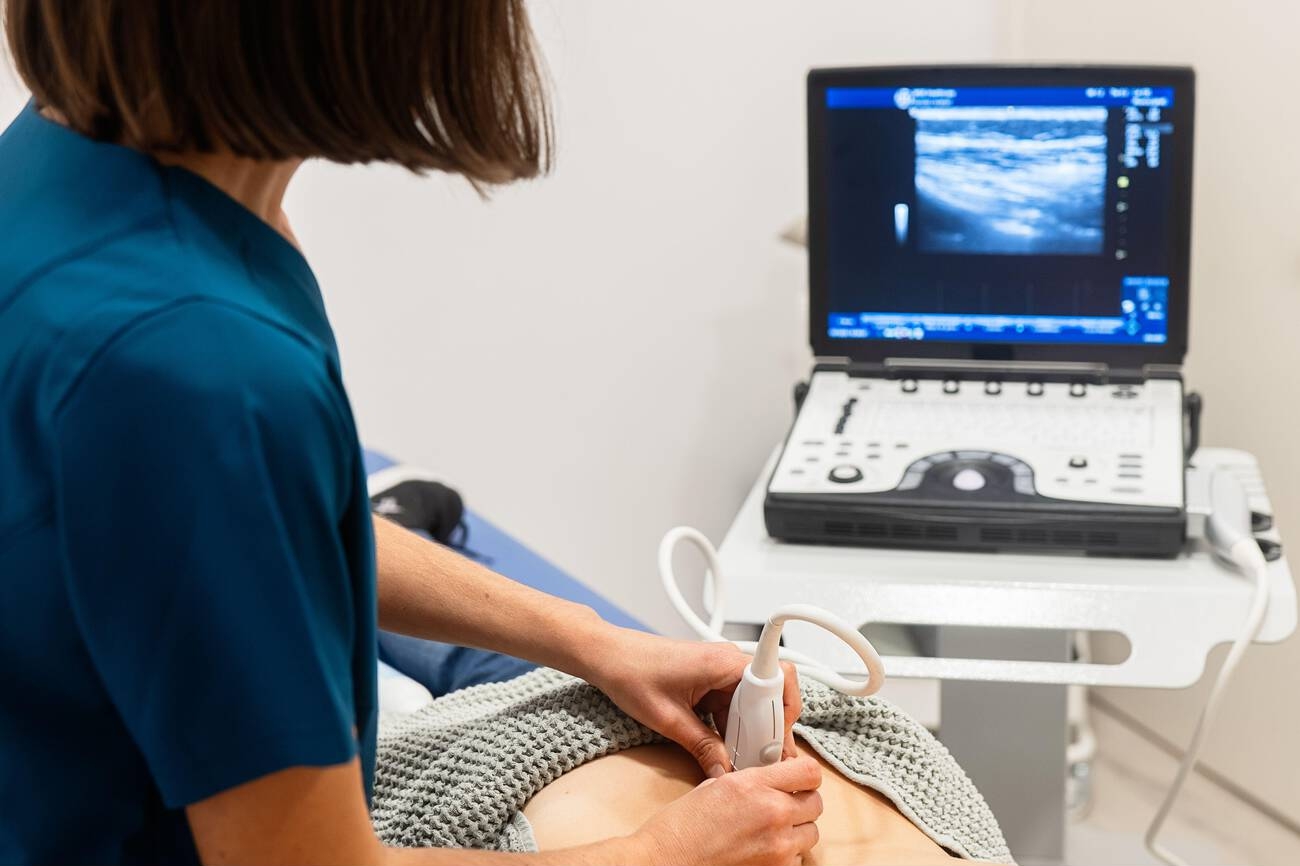
A curved probe at work on the abdomen, here on a conventional cart system. The probe in the hand is one configuration, a single head matched to deep scanning; a wireless handheld makes the same choice in a head that runs from a phone. Image: Goleisureintl, CC BY 4.0.
The trouble is set in physics, well before any marketing. A single crystal can reach deep or it can draw fine. It cannot do both at once. The heart wants a third thing entirely, a small face that slips between two ribs and sweeps a fan across the chest. A probe that claims all three has found one of a few ways around a hard wall. Two numbers describe the wall. Sound loses strength as it travels, and it loses it faster the higher its frequency runs, roughly a decibel for each centimetre and each megahertz of pitch. A three-megahertz wave still carries through twenty centimetres of liver. Push the pitch to fifteen and the same wave is spent inside five. Detail runs the opposite way along that curve: the higher the pitch, the finer the grain it draws. A crystal is tuned to one end of the curve only. The same physics fixes how fine a picture the probe can draw. A higher pitch packs more wave cycles into each millimetre, so it separates two points lying close together. The width of the beam counts as much: a tight beam tells apart two structures lying side by side. This is the trade behind every configuration on the market, and no engineering erases it. Every handheld hides it behind presets, a button that loads the depth, the frequency, and the gain an exam wants in one tap.
A clinic can own a separate head for each job. It can carry two heads fused into one body. It can buy a single head clever enough to play all three parts. None of the three comes free. None is best for everyone. Each is a different bargain. The right one depends on the work a clinic actually does.
Own a head for every job
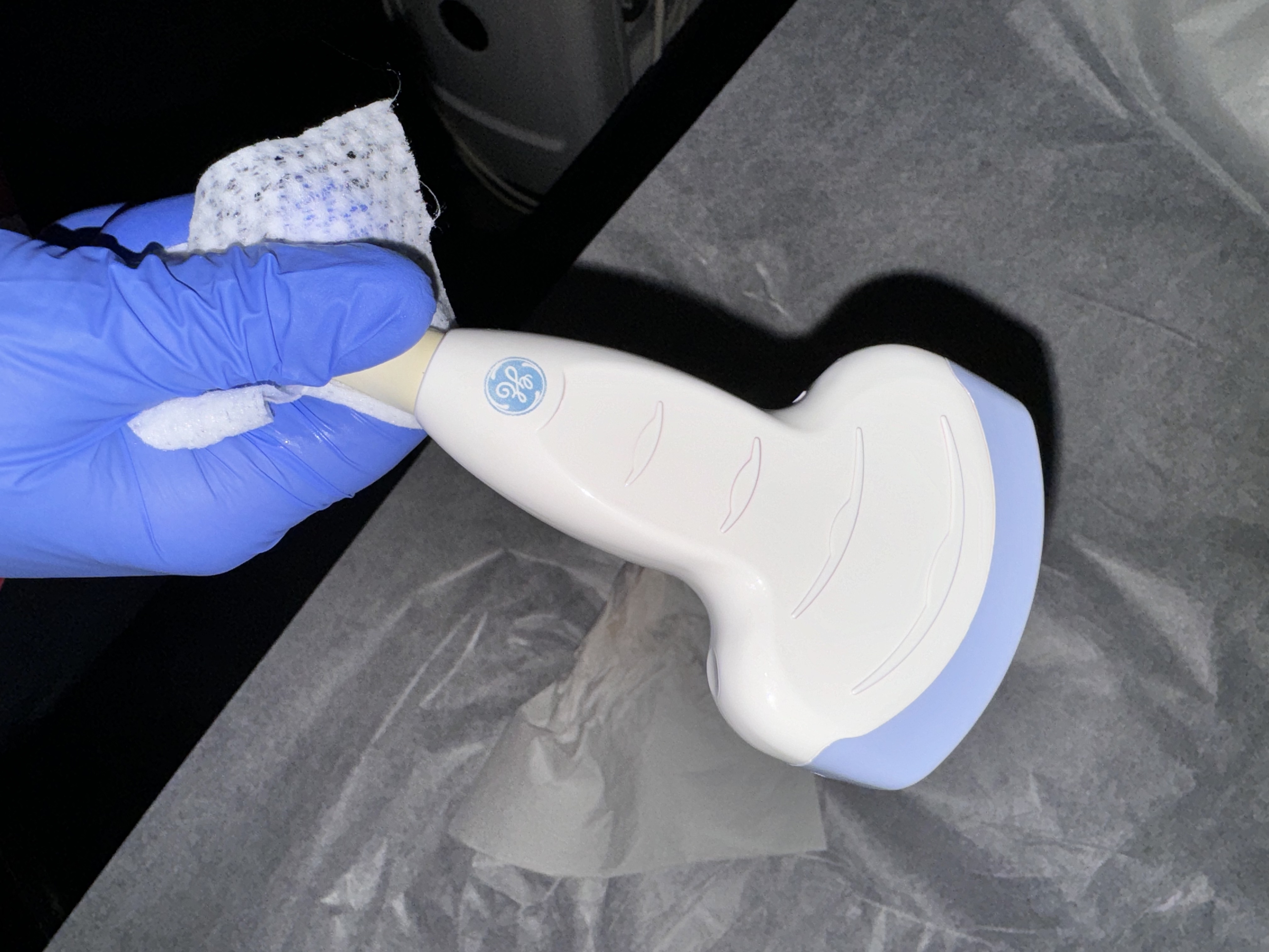
A single ultrasound scan head, wiped down between patients. This is one probe for one kind of exam, the sort a clinic either owns a set of, one head to a job, or replaces with a single broadband probe that covers them all. Image: Harrison Keely, CC BY 4.0.
The oldest answer is also the easiest to picture: buy a head for each thing you scan. A convex head for the belly, a linear head for the surface, a phased head for the heart, every one of them built to do its single job as well as the craft allows. Each head is a pure specialist, no part of it bent toward a second job. The picture, in every role, is the sharpest a probe can make. The three heads are genuinely different instruments. The convex head runs at two to five megahertz over a curved face that fans a field deep into the belly and the pregnancy. The linear head runs at seven to fifteen megahertz and higher over a flat face that holds fine detail across the first few centimetres. The phased head runs low again, two to four megahertz, on a tiny footprint that aims a sector beam through the gap between two ribs at the heart. Put any one of the three in another’s place and it gives up much of what makes it good.
Weighing that full set of single heads against one probe that tries to do everything is the first fork a buyer reaches. Owning the set means owning the baggage. Three or four probes are three or four things to charge, to clean, to keep track of, and to reach for when the exam in front of you changes.
A full set is also a real outlay. Three or four dedicated probes cost more together than one broadband head, each carries its own warranty and its own replacement price, and a clinic that breaks or loses one buys a whole probe to replace it. How much that outlay stings comes down to how often the probes earn. A unit that scans all day spreads it across thousands of studies, down to pennies a scan. Idle in a drawer much of the week, the same hardware is money sitting still, depreciating month after month.
The baggage bites hardest on the move. Carried between wards and villages, every probe in the bag is one more thing to charge and one more thing to lose, and a head that never comes off starts to look like the better trade. The set does fail gracefully. When one head dies, the others carry on and the clinic keeps scanning.
Fold two heads into one
The next answer trims the baggage without surrendering the dedicated crystals. Two transducers built into one probe sit back to back, or side by side under a single cover, and a roll of the wrist brings up whichever face the exam needs. One probe charges once and does two jobs. Nothing ever leaves the hand. The two crystals are real, separate arrays, each tuned to its own band, packed nose to nose with the electronics shared between them. That sharing is the engineering trick and the source of the extra bulk: one battery, one radio, one processor feeding two faces.
Which two faces get fused is a bet on the work. A phased face paired with a linear one serves the clinician who reads a heart and then drops a line into a neck without setting the probe down. A convex paired with a linear serves the one who works the belly and the surface by turns. The two crystals share a handle, a battery, and a radio, so the switch costs nothing but a twist. The fused probe is a niche tool sold to a known market. The phased-and-linear pairing goes to emergency and critical care, where one hand reads a heart and then guides a needle into a vessel. The convex-and-linear pairing goes to general and travelling clinics, where a belly scan and a surface scan follow one another all day. Some makers reach the same end with stacked crystal layers behind a single face, firing one band or the other from a more complex array, with no flip at all.
Two transducers under one shell run heavier and longer than a single head. The idle face is dead weight until its turn comes. Roll the wrist twenty times a shift and the heft earns itself back many times over. Live in one kind of exam and it is weight carried for nothing. The dual head suits the clinician whose day genuinely straddles two territories, and that clinician alone.
One head for everything
The boldest answer refuses the choice. A single broadband head that plays convex, linear, and cardiac in turn runs across a wide spread of pitches, dropping low for the belly, climbing for the skin, narrowing to a fan for the heart. Nothing detaches. Nothing flips. A tap on the screen changes the probe’s whole character. One small body in a coat pocket holds the entire range, charged once, with nothing to swap and nothing to drop. It is the closest the handheld has come to its founding promise, a whole imaging room shrunk to a thing on the end of a phone. The trick inside is a wide-bandwidth crystal, a single array built to resonate cleanly across a far broader span of frequencies than an ordinary head. The software drives it low for a deep preset, high for a shallow one, and steers it into a sector for the heart, all from the same hardware. A modern broadband array can hold a usable picture from around two megahertz to past twelve, which is the bulk of the range three separate heads would cover. Bandwidth, channel count, and processing all have to match. All three have to be strong together, or the headline number gives back less than it promises.
The price is paid quietly, in the picture. A crystal asked to do every job is never quite the equal of one that does a single role, cleanest in the middle of its range and softening a shade toward the far edges. For nearly every bedside question that softness never surfaces; the answer lands long before fine grain would matter. The one study it struggles with is the one that turns on the finest texture of a small thing, where a dedicated head still pulls ahead. In daily use a good broadband head holds a clean picture across the whole of an everyday workload. It reads a gallbladder, a lung, a bladder, a vessel, and a focused heart from the one body, switching at a tap. The soft edge shows on the studies that lean on fine grain: a small thyroid nodule, a shallow tendon, the early texture a dedicated linear head would render a notch crisper. Meet those studies rarely, and the trade is an easy one to make. The single head is also the simplest to teach and to support, one probe and one set of presets for a whole staff to learn.
The fine print that decides it
Within any of these covers, the same small print decides whether a probe honours its bargain. The first line is the count of elements, the slivers of crystal that throw the sound out and gather it back. How many elements a head carries sets how tight a beam it can draw: a head of a hundred and ninety-two picks out detail that one of a hundred and twenty-eight smears together. The number means little until two probes meet the same patient, where the finer array lifts a clean edge out of a blur. More elements pull more current, throw more heat, and lean harder on the processor behind them, so a handheld settles on a count its battery and its grip can bear. A clinic rarely counts channels at all, and reads the result as how deep the picture stays sharp and how cleanly a small structure comes through. The count drives more than sharpness. A denser array steers and focuses its beam with finer control, holds focus deeper into the field, and runs the live measurement packages a clinic leans on. A jump from a hundred and twenty-eight channels to a hundred and ninety-two, or to two hundred and fifty-six, earns its keep on a deep abdomen and a moving heart, where the beam has to work hardest. There is a floor below which more channels stop paying back. Past a point the beam is already as tight as the crystal and the tissue allow, and extra channels add only data and heat. The sweet spot for a pocket probe sits where the picture is good enough for the job and the probe still runs cool and long on one charge.
The second line is the join. On a probe with clip-on heads, the connector that locks a head to its handle carries every element’s signal across a contact that has to seat true ten thousand times, shrug off the gel and the wash-water between patients, and let go in an instant for the next head. A loose or dirty connector picks the worst moment to show as snow across the screen, or as no screen at all. A single bent or oxidised pin drops out one strip of the image, a fault a clinic learns to read as the probe, not the patient. A fully sealed wireless probe does away with the join, and pays for that in separate bodies to own and to charge. A clip-on connector is a dense block of gold-plated pins, one or more for every channel in the array, sprung to seat under a firm push and latched so a busy hand cannot drag it loose. It has to stay watertight through a gel-and-alcohol life and survive thousands of swaps with its pins straight and its contacts clean. The makers who build the connector as a precision part are the ones whose probes still read clean after a year on a ward. The other thing a buyer cannot see is how a probe ages. A crystal loses a little output over years, a battery holds less charge, a connector wears down over thousands of swaps. Build quality decides how a probe ages. A weakly built one can fail in its second year, out of warranty, with no part a clinic can replace. What the probe is still good for three years in rides on all of this, none of it printed on a box.
The last line matters only in a hurry. Whether a head change interrupts the scan or rides straight through it stays invisible on a spec sheet until a cardiac arrest makes it the only thing that matters. A swap that drops the image and reloads the software burns the seconds a crashing patient cannot spare. A hot swap keeps the study live, the new head waking exactly where the old one stopped, the reader’s eyes never leaving the patient. Under the surface it means the app holds the patient record, the measurements, and the exam open through the brief moment the probe hardware needs to re-enumerate, a few hundred milliseconds the reader never feels. Some systems rebuild the whole session on each change, costing ten or twenty seconds, the difference between a tool used in a code and one left on the shelf.
Running it from a phone
A wireless probe is a whole machine in a sealed shell. Inside sit the array, the beamformer, a battery, a processor, and a radio. The phone or tablet is a screen and a set of controls, not the engine. The probe does the heavy work of forming the image and sends a finished picture over the radio link, which is why a modest phone can run an expensive probe. The processing inside the probe is what makes the wireless link possible at all, since a finished frame is small enough to send where the raw data would choke the radio.
That model sets the practical limits. The battery lasts a working session, an hour or two of live scanning, and charges between patients on a cradle or a cable. The radio link has to carry a moving image with no stutter, so the probe and the app are tuned to each other, one reason a probe and its software come as a pair. Heat builds in the sealed shell through a long scan, and the probe throttles itself before it grows warm in the hand.
None of this reaches the screen, and that is the design. A clinician sees a live picture and a few buttons. Fitting the array, the beamformer, the battery, the heat budget, and the radio into a thing the size of a thick marker, run from a phone, is what every configuration choice sits on top of.
The link also shapes the workflow. A study lives on the phone, and the phone moves it: into the patient record, up to a colleague across the building, out to a specialist in another city for a second read. How freely it moves depends on the app, and a closed one strands the images on the device, out of reach of the systems a clinic already runs. Battery life is a constraint a spec sheet understates. A probe rated for an hour of continuous scanning lasts a morning of stop-start use, and a clinic that scans back to back keeps a second probe or a charging cradle within reach, since a flat probe halfway through a clinic stops the clinic cold. A spare battery or two keeps a long list moving with no pause for the cradle.
Choosing, and changing your mind
No configuration is right for everyone, whatever each maker’s brochure implies. The set goes to the busy department that wants the best of every picture and never has to leave the room to get it. The broadband head goes to the mobile service that values one light thing it never has to swap. The dual head goes to the team whose day is split clean down the middle. The honest way to choose is to put the brochures down for an afternoon and put the probe in your own hand, on your own patients. An hour of that teaches more than any sheet of numbers. The probe is only half of what a clinic buys. The software around it, the way it stores and shares a study, the speed of its updates, the length of its warranty, and the price of a replacement head years from now all ride on the same purchase. A cheap probe tied to a thin, slow app is a poor deal at any price. Two plain questions cut through the sales talk: what does this clinic scan in a typical week, and who carries the probe. Run the same four exams all day, and a set earns its place. Carry ultrasound from room to room all shift, and one head earns its place. The trial matters because no two clinics scan alike. A broadband probe that suits a GP running focused scans can fall short for a sonographer chasing fine detail all day. Two clinics put the same probe to different work, and matching that work is the buyer’s real task. The surest test of an app is mundane: scan a patient, save the study, and send it where it has to go, then see how many taps that took. A week of the clinic’s own logbook decides it better than a day of demonstrations, naming the exams that actually fill the schedule.
The choice is not for life, either. A clinic can start with one broadband head and add a dedicated probe later, when its work narrows or its budget loosens. A modular base also survives a maker’s stumbles, since one discontinued head leaves the base and the rest of the set working. The market moves fast enough that the head leading this year may trail in three, which rewards a base a clinic can re-head cheaply. Buy for the work in front of you, leave a little room for the work coming, and the right configuration mostly disappears into the day, never thought about again.
Common questions about handheld probe configuration
What is an interchangeable-probe ultrasound system?
A handheld that covers many exams by changing its probe. A base accepts a set of single-purpose heads, one swapped for the next to suit the exam: a convex head for the deep belly, a linear head for the surface, a phased head for the heart. The other path is a single broadband probe that does all of it with no swap.
Is one multi-purpose probe better than a set of single probes?
Neither beats the other outright. The broadband probe is lighter, charges once, and asks for no swap. The set draws a sharper picture in each role, since every head is a specialist. Which one fits comes down to the work and who carries it through a shift.
What is a dual-head ultrasound probe?
One probe holding two transducers in a single body, commonly a convex face with a linear face, or a phased face with a linear one. A turn of the wrist brings up the other face, with no head to detach. One probe does two jobs, a little larger and heavier for it.
Do more transducer elements mean a better image?
Up to a point. More elements tighten the beam and sharpen the picture, so a 192-element head resolves what a 128-element head blurs, and each extra element adds cost and heat, so a handheld picks a count that serves the job without cooking the probe in the hand.
What is hot-swapping a probe?
Changing the head without stopping the scan. The session stays live, and the new head picks up exactly where the old one left off, with no reload. In an emergency, that unbroken flow can decide whether the scan gets used at all.
About the Author
Julien Mercier
Senior R&D Engineer · Medical Ultrasound Transducer Development
Senior R&D Engineer with an M.S. in Applied Physics and over 15 years of experience in medical ultrasound transducer development, specializing in the design verification and performance testing of high-frequency imaging transducers. Currently leading the development and verification of the company’s next-generation high-frequency linear-array transducer, responsible for imaging performance evaluation and reliability analysis in preclinical testing. Brings extensive hands-on experience in piezoelectric element tuning, beamforming parameter optimization, and system-level performance testing.