


































Veterinary Ultrasound Handheld Wireless Probe For Animals
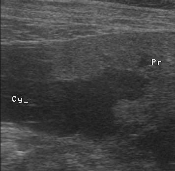
Veterinary ultrasound is the reading of an animal’s insides through a handheld probe, the same scan run on a kitten in a clinic or a cow standing in a barn. One pocket-sized machine covers that whole range. What changes from a cat to a horse is the depth the sound reaches, the window it works through, and the place the work happens.
One probe, the whole animal kingdom
The span is the thing that sets veterinary work apart from the human kind. The body under the probe might weigh two kilograms or seven hundred. It might be buried in dense fur, balanced on three legs, pregnant with eight at once, or kicking at the person holding it. The room is often a barn, a muddy field, or the back of a car on a farm round. One probe has to find a usable picture through all of that, which is why the operator carries the weight of the work. The pocket scanner is the easy half. Knowing where to lay it on a creature nobody demonstrated for you is the craft that takes the years. The same probe turns up in stranger places than a clinic or a farm: a zoo vet checks a pregnant big cat through the bars, a wildlife team scans a sedated deer in a field, a researcher follows the same lab animal week by week. The reach of the tool is, in the end, the reach of the animal kingdom itself.
What changes from a cat to a horse
Depth is the first thing that shifts between one animal and the next. A kitten’s bladder sits a centimetre under the skin and wants a high-frequency probe that draws fine, shallow detail. A cow’s pregnant uterus lies an arm’s length inside the rectum. A horse’s tendon runs deep down the back of the leg. Those deeper targets want a lower frequency that trades fine detail for the reach to get to them. A linear probe runs roughly in the seven to fifteen megahertz band, sharp for the first few centimetres of depth. A convex probe drops to around two to five megahertz, reaching fifteen to twenty centimetres or more into a large animal. That trade between detail and depth is fixed by the physics of sound. It is the reason no one frequency suits both a ferret and a bull.
The probe comes in a few shapes to match. A flat linear probe, high in frequency, reads tendons, eyes, and the shallow belly of a small dog. A curved convex probe, lower in frequency, fans the beam out wide, reaching deep enough for an abdomen or a late pregnancy. A small, tightly curved microconvex probe fits between ribs or into a small body.
For farm breeding work there is a long rectal probe built to ride along the arm. A handheld system covers the whole range one of two ways, with heads that clip on and off, or with a single broadband probe that shifts its frequency in software across the whole range.
The species also sets the window, the path the sound is allowed to take. Gas and bone stop ultrasound dead. Animals carry both in awkward places. A dog’s gas-filled gut hides the pancreas behind a grey smear. A horse’s lung walls the heart off to a narrow slot between two ribs. The scan works around these by choosing where to sit the probe and how to tilt it.
The probe reads movement as well as shape. Colour Doppler paints blood flow onto the grey picture, so the flow lost inside a twisted testicle, the anger of a tumour, or a flickering fetal heart all show up live on the screen. On a patient that cannot point to a symptom, watching how the tissue moves is one of the few ways its body gets to speak.
Reading the grey picture
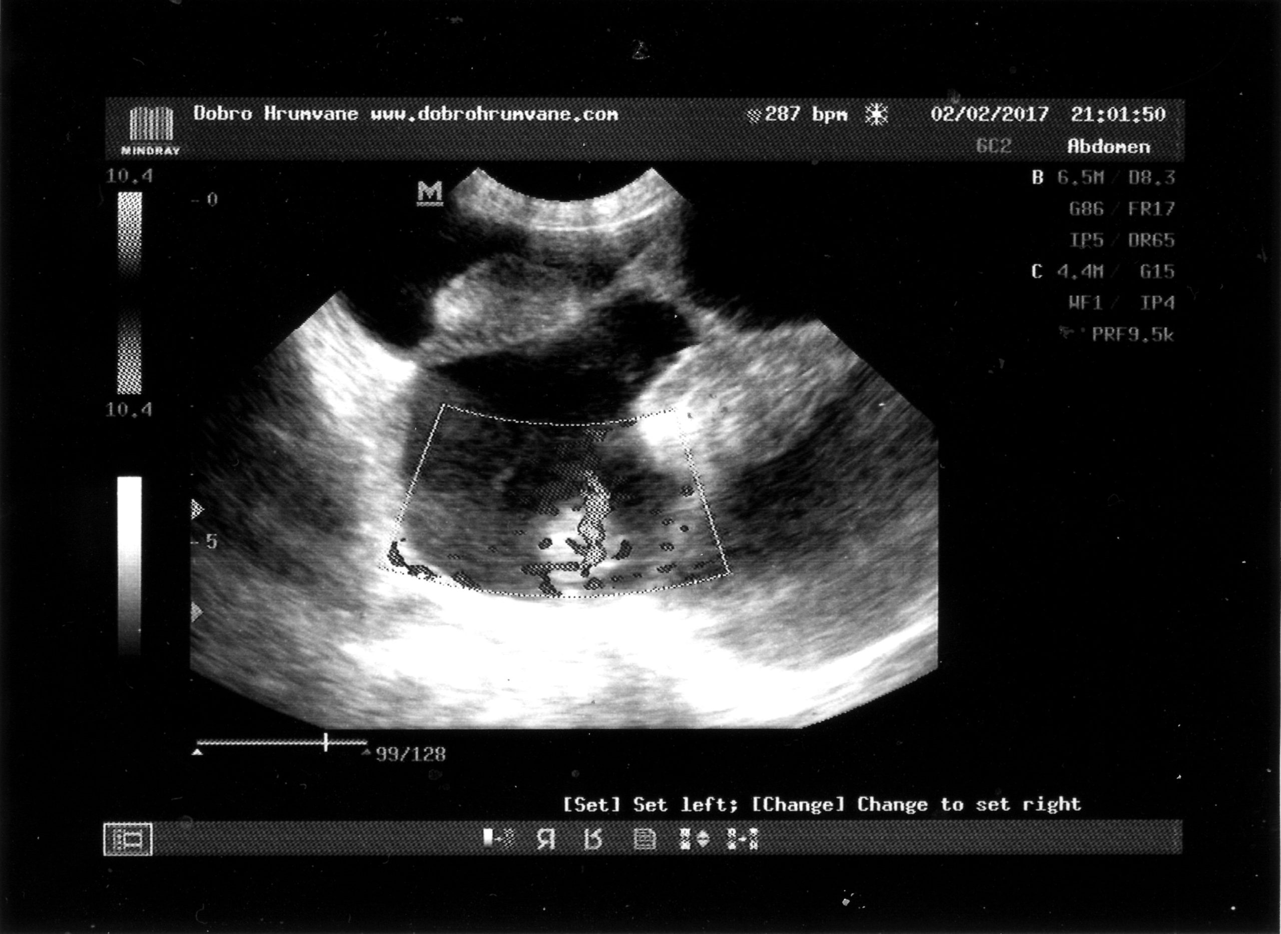
The image the probe draws is a map of echoes. Sound sent into the body bounces back wherever the tissue changes. The machine paints each return as a dot of grey, its shade set by the strength of the bounce. Fluid lets the sound straight through, so a full bladder or a pocket of blood reads as clean black. A stone or a slab of bone throws the sound right back and drags a dark shadow out behind it. The soft organs sit in the middle of the scale, each with its own texture that a trained eye learns as that organ’s normal.
Reading the picture is a matter of knowing that normal and catching where it breaks. A kidney holds a clear line between its outer rind and its inner core until disease rubs that line away. The operator works by comparison, reading one side of the body against the other, or the sick animal against the memory of a hundred healthy ones. None of this is automatic. The same grey patch can be read as nothing or as everything depending on the eye behind the probe.
A second trap is the artefact, the mark on the screen that no real tissue put there. Sound bounced twice between two strong surfaces can draw a structure deeper than it sits. A smooth curved wall can throw a bright tail or a dark edge that a beginner reads as disease. Part of learning the grey picture is learning which of its marks belong to the animal and which belong to the machine.
Setting the machine for the animal
Before the picture means anything, a few controls have to fit the patient in front of the probe. Depth sets how far down the screen reaches, matched to where the target organ sits. Getting it wrong either buries the target off the bottom edge or shrinks it to a speck up top.
Gain sets the overall brightness, lifting a dim picture out of a thick-coated dog or pulling the glare back on a thin one. A focus marker tells the machine which depth to sharpen, parked at the level the organ sits. Many handheld systems fold all of this into species presets, so a single tap for small-animal abdomen or equine tendon loads a sensible starting point that the operator then trims.
The last setting is the one between the probe and the skin. Air is the enemy, since sound will not cross it, so the join has to be airtight. A patch clipped to the skin and flooded with warm gel gives the beam a clean road in. On a farm, where shaving a whole flank is not always on offer, alcohol poured straight onto the coat can wet it down enough for a fast look. A picture that comes up grey and grainy is as often a coupling fault as a machine one. A fresh blob of gel rescues more bad images than any dial on the screen.
A frozen frame turns the live scan into a record. The operator stops the image at the right moment, drops a pair of calipers on the screen, and reads off a measurement, the length of a kidney, the depth of a pocket of fluid, the diameter of a tendon. Those numbers go into the notes and become the baseline a later scan is judged against. A saved still also travels better than a memory, passed to a colleague or filed as proof of what the screen held.
Guiding the needle
The probe does more than look. It steers a needle. With the tip tracked live on the screen, a vet draws a sample from a swollen node, drains a pocket of fluid from a chest, or places a catheter into a vein no finger can reach. The same guidance works across the size range, from a cat’s bladder up to a mare’s abdomen. On a farm it sets a biopsy into a horse’s tendon, or puts a needle in the right spot before a joint is injected. Watching the tip the whole way in turns a blind stick into a precise one, and pulls a diagnosis from a few cells.
The machine is the easy part
Learning to read what a pocket scanner shows takes years.
The questions that recur in every species
Strip away the species and a short list of questions comes back again and again. Is she pregnant, and is the litter or the foal alive. Is the heart squeezing or failing. Is there fluid where there should be none, blood in a belly or water around a lung. Is a tendon torn, a kidney blocked, a gut wall too thick. The same handful of questions drives the scan whether the patient is a cat or a cow. That is the deeper reason one probe can serve them all.
What changes between species is the route to the answer and the look of normal. A pregnancy is read through the belly wall in a dog, through the rectum in a mare, each time the same search for a sac and a heartbeat. That shared core is why a vet who learns to read one species moves to the next by learning a new anatomy, and why the skill, once won, carries across the whole range of animals.
Those recurring questions are also where the money sits. A pregnancy confirmed early lets a breeder cull the open females before a winter of feed. Grading a heart in the room saves a referral fee. Finding a bleed in the minutes after a road accident decides surgery while it can still help. The scan holds its place in animal work through how often the same few answers change what happens next.
Three kinds of work
The field sorts itself by the size of the animal and the place the work has to happen. The clinic side is small-animal work on dogs and cats, the busiest corner by a wide margin, done on a table with a small patient lying close to the probe. The detail of that work, organ by organ, sits on its own page.
The farm side is large-animal ultrasound on horses, cattle, and sheep. Here the machine travels to the patient. The scan reaches the organ that counts by the shortest safe route, which more often than not runs through the rectum.
A third strand stands apart from any single species. Veterinary scanning has its own standards and training, the bodies, courses, and named protocols that set out what a proper exam looks like and how it gets recorded. Each of these three opens onto its own page below.
The animal does not hold still
The deepest break from human scanning is simple. The patient does not cooperate. No animal holds its breath on cue, lies flat to open a window, or says where it hurts. A frightened dog clenches its belly into a board over the organ being hunted. A horse leans away the instant the probe meets a sore leg. Every picture has to be caught in the gaps between one movement and the next. The person scanning reads the animal’s body at the same time as the screen.
Restraint does the job a spoken instruction does for a person. A nurse cradles the dog, a halter steadies the horse’s head, a chute pins the cow so she cannot swing. Hair is the next fight, since fur traps a layer of sound-blocking air against the skin, so a patch is clipped and soaked with gel before the probe reads anything clean. Where a clip is refused, a heavy soak and a patient hand are the fallback.
When restraint and a calm hand fall short, a light sedation buys the stillness the scan needs, dropping a fractious cat or a kicking horse into a state quiet enough to read. It is a balance, since a heavily sedated animal can shift the thing being measured, slowing a heart or slackening a gut, so the dose is held to the least that keeps the patient still.
Cutting the cord
In a barn a cord turns into a real problem. The older farm scanners hung in a box on the operator’s neck, wired down to a probe, with a screen strapped wherever the eyes could reach. A wireless probe carries none of it.
The probe talks to a phone or a tablet over its own signal, so the screen becomes whatever the operator props on a rail or wears on an arm. A hand comes free. The dangling gear that used to snag on a chute or a swishing tail is gone. The probe holds its own battery and electronics, sealed against gel, dung, and the wash-down that ends a dirty scan.
Going wireless also frees where the picture gets read and what happens to it after. A loop saved on the phone drops into the patient’s record and travels down the line to a specialist for a second read, which can stand in for a long drive to a referral centre. For a vet on a farm round, the whole imaging kit shrinks to a probe in one pocket and the phone already in the other.
A single charge runs long enough for a morning of farm calls or a clinic list, after which the probe sits on a pad to fill again. The sealed body shrugs off the wash-down, the splash of dung, and the knock of a drop onto a concrete floor, the daily abuse a working barn hands out. Stripping out the cable also strips out the part that fails first, since a flexed wire and a plugged socket are where an older scanner usually died.
From cart to pocket
For much of its history, ultrasound on animals lived on a wheeled machine the size of a fridge, parked in a university hospital or a referral practice. A farmer or a first-opinion vet who needed a scan booked it, drove to it, and waited. The access was poor. The cost of a machine put one of your own out of reach for an ordinary clinic.
The pocket probe turned that around by separating the expensive thinking from the part that touches the animal. The processing that once filled a cabinet now sits in a head the size of a bar of soap, talking to a phone the clinic already owns. The price of getting started fell far enough that a single-vet practice or a roving farm vet could carry one. The scan spread into ordinary clinics and out onto the farms.
The shift also changed who carries the cost and who gets the picture. A clinic no longer buys one shared machine and rations it across the staff, since a probe is cheap enough that several can live in one practice, one in each consult room or one in each vet’s bag. On a farm the sums are starker still, because a scanner that covers its cost in a single season of accurate pregnancy diagnosis is an easy buy for a breeder to sign off.
Sound that does no harm
One reason the scan gets reached for so freely is that it does no damage. Ultrasound carries no radiation. It works by sound alone, so there is no dose to add up and no shield to stand behind. A pregnant animal can be scanned again and again across her term, safely for the litter, which is what makes it the tool of choice for following a pregnancy as it unfolds. The same loop can be run a dozen times in a workup.
The machine still puts energy into tissue. Two small numbers on the screen, a thermal index and a mechanical index, track how much of it. In animal work the levels used sit well inside the safe range. The operator keeps them as low as the picture allows, turning the power down once the image is good enough. That freedom to scan often, without counting exposures, is a quiet part of why the probe became an everyday instrument.
What it hands to other tools
Ultrasound has hard edges. Sound bounces off gas and bone, so the inside of an air-filled lung, a gut packed with gas, and the marrow of a bone stay closed to it. A broken leg is still a job for X-ray. A lung gives up only its surface, read through the pattern of echoes coming off that surface.
Reach is the other wall. On the largest animals a pocket probe runs out of penetration before the sound gets through the depth of a draught horse’s belly or across to the far side of a bull’s chest. The picture fades to grey noise at that depth. A scan that needs that depth, or a study that turns on the finest detail, is where the handheld hands the case on to a bigger machine.
Ultrasound joins the older tools. An X-ray still rules the skeleton and the gas-filled chest, where sound cannot follow. A blood test names what a swollen organ cannot. At a referral hospital, a CT or an MRI builds a picture in depths and planes a handheld probe never reaches. The pocket scanner does its work by answering the first and commonest questions on the spot, then handing the harder ones up the line.
The picture also leans hard on the hand that takes it. The same loop of bowel can look normal or alarming with a change of angle or a turn of a dial. That dependence is why the training and the standard count for as much as the device. A scanner answers the question it is pointed at.
The breadth of one probe
A single probe, run by one person, covers a long list of everyday jobs across the animal world. The work runs from confirming a pregnancy and counting the young, to reading a heart for the diseases of a cardiology day, to finding a stone in a bladder or fluid in a belly or a tear in a tendon. It takes in the fast sweep for a bleed after a road accident, the guided needle into a node, the drain into a fluid-filled chest, the check on a kidney in slow decline, the look at an eye behind a cloudy cornea. One pocket scanner reaches all of it. The same grey picture, read by a trained eye, answers each of these from the outside of an animal that never has to be opened up. That breadth, in a tool light enough to carry and cheap enough to own, is what put ultrasound into the daily round of veterinary work across every kind of animal there is.
The tool that goes anywhere
Across that whole range, the scanner stays one steady tool. The animal, the setting, and the hands around it are what move.
Common questions about veterinary handheld ultrasound
Can a handheld wireless probe scan animals as large as cattle and horses?
Yes, within limits. A handheld probe reaches the targets a farm scan aims at, the reproductive tract through the rectum and the tendons down the leg, because those sit within its depth. The deepest scans on the largest animals are where a sealed pocket probe can run short of penetration. A cart-based machine still holds its place there.
Does the animal have to be shaved before a scan?
Usually a small patch. Hair traps a layer of sound-blocking air, so a window is clipped and soaked with gel or alcohol. A heavy soak can sometimes get a usable picture without a clip, especially on a thin-haired belly.
Is one probe enough for every kind of animal?
It depends on the spread of work. One probe handles a clinic of cats and dogs. Reaching from a kitten’s bladder up to a deep bovine pregnancy takes either a probe swap or a broadband probe that shifts frequency across that span.
How does a pocket scanner compare with a full ultrasound machine?
For the focused questions of field and clinic work it is generally close enough to act on. The hardest studies, turning on the finest detail or the deepest reach, still go to a cart-based system. The handheld gives up a little image quality for the freedom to go anywhere, cordless, on a battery.
Who uses handheld ultrasound on animals?
A widening group, from a single-vet rural clinic to a city hospital with a probe in every consult room, plus mobile vets working out of a car and farm vets going herd to herd. A cheaper, pocket-sized probe lowered the cost of owning one.

About the Author
Julien Mercier
Senior R&D Engineer · Medical Ultrasound Transducer Development
Senior R&D Engineer with an M.S. in Applied Physics and over 15 years of experience in medical ultrasound transducer development, specializing in the design verification and performance testing of high-frequency imaging transducers. Currently leading the development and verification of the company’s next-generation high-frequency linear-array transducer, responsible for imaging performance evaluation and reliability analysis in preclinical testing. Brings extensive hands-on experience in piezoelectric element tuning, beamforming parameter optimization, and system-level performance testing.