


































Optic Nerve Sheath Diameter ONSD Measurement Handheld Ultrasound
The optic nerve runs from the back of the eye to the brain, wrapped in a sheath that carries the same fluid that bathes the brain itself. When the pressure inside the skull rises, that fluid pushes outward and swells the sheath, most of all just behind the eye. The width of the sheath there, the optic nerve sheath diameter, is a number a handheld ultrasound can read in a minute, through the closed lid, with no needle and no scan of the head. It is a window onto the pressure in the skull, opened from the front of the eye.
A pressure gauge behind the eye
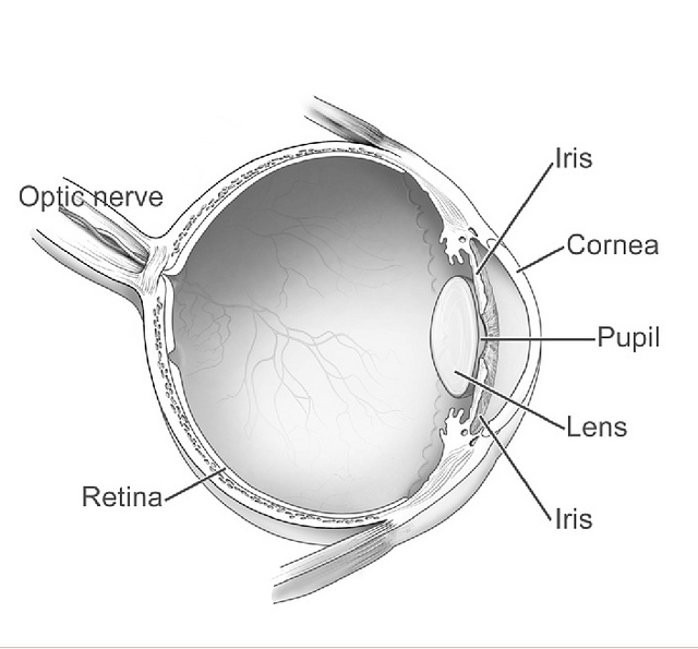
The eye and the brain share a covering. The optic nerve travels to the brain inside a sleeve of the same tough membrane that wraps the brain, the dura. That sleeve is built of the three layers that wrap the brain itself: a tough outer dura, a web-like arachnoid, and a thin pia laid against the nerve. Between them runs the subarachnoid space, the channel the cerebrospinal fluid moves in. The cuff of fluid around the nerve is continuous with the fluid around the brain through the back of the eye socket. The pressure in one is the pressure in the other. In that sense the optic nerve is a length of the brain reaching out to the eye. It carries the brain’s own pressure with it. The nerve takes a gentle slack curve through the orbit, a little longer than the straight path, so the eye can turn without tugging on it.
So a rise in pressure inside the skull does not stay inside the skull. It travels down the fluid in the sheath to the back of the eye. The front stretch of the sheath, the part just behind the globe, is the most elastic length of it. The fine strands that tether the nerve to the sheath are sparsest in that front stretch, which is what lets it balloon there. The sheath is loosest about three millimetres behind the globe, where its wall gives most under load. It is there that the rising pressure shows first, ballooning the sheath outward. A swollen sheath behind the eye is the footprint of a swollen pressure in the head. The sheath answers a change in pressure within minutes. It settles back when the pressure falls, which lets the same scan follow how the pressure moves over the hours. A pressure that has stayed high for weeks can leave the sheath stretched even after it eases, a wider baseline the history explains. In a long-standing condition like idiopathic intracranial hypertension, the sheath can sit wide for months while the pressure stays up.
Measuring that swelling is what the scan does. The fluid-filled eye is a clear acoustic window, the one place on the head where sound passes freely to the soft tissue behind it. Ultrasound reaches the sheath through the soft eye without a cut, measures its width to a fraction of a millimetre, and does it again as often as needed. Ultrasound studies in the 1990s mapped how the sheath swells when the pressure behind it rises. The bedside measurement grew out of that work into emergency rooms and intensive care units. The measurement is taken on a B-scan, the two-dimensional picture, where the nerve and its sheath stand out as a clear band against the orbit. The number it gives stands in for a pressure that otherwise takes a drill and a bolt through the skull, or a needle in the spine, to take directly. Neither of those can be done in a hallway in a minute. A widening sheath is an early, gentle warning of a danger building out of sight, read off the one soft part of the head a probe can reach.
Measuring the sheath
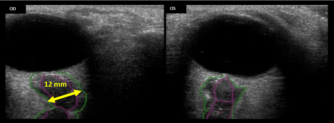
The scan is done over a closed, gel-covered lid, the gentlest touch the eye allows. The probe never presses; a pressed eye gives its own false numbers. A clean, single-use gel over the closed lid carries the beam in. The closed lid keeps the eye surface untouched. A drop of gel and a light hand are all the eye needs: no drops, no dilation, no pressure on the globe. Position counts for the number too. The patient lies flat with the head neutral, since lifting the head drains a little pressure and narrows the sheath. The patient looks straight ahead under the lid and holds the eye still. The beam aims through the dark circle of the eye to the structure running away from its back wall. That structure is the optic nerve and its sheath, a band reaching back into the orbit toward the brain.
On the screen the nerve and sheath show as a band leaving the back of the globe, running down and away into the grey of the orbit. The nerve runs straight back from the disc in a healthy eye, a steady dark band the calipers can sit across. The two together, the nerve at the core and its sheath around it, make the width that is measured. When the pressure is high, a thin dark crescent of fluid can show in the sheath around the nerve, the fluid that has backed up from the brain. The optic disc itself can bulge forward into the back of the globe, a second sign of the same trouble. The same probe in the same sitting can sweep the rest of the eye, the retina, the vitreous, the lens, so the sheath reading comes as one part of a fuller look.
The width is taken about three millimetres behind the globe, at a set depth along the nerve. The three-millimetre rule comes from early ultrasound studies of the sheath, which found that depth gave the steadiest, most repeatable number. That depth is where the sheath gives most under pressure, the spot that swells soonest and furthest, and the spot where the screen draws the edges most clearly. The clinician places the calipers across the full sheath at that depth, edge to edge, square to the run of the nerve. The two bright walls of the sheath are the edges the calipers land on, one on each side of the nerve. The width counted runs from the outer edge of the sheath on one side to the outer edge on the other, taking in the nerve and its fluid cuff together. The number comes up in millimetres on the screen. A second look in a plane turned ninety degrees confirms the band was caught across its true width. Turning the eye out by about thirty degrees stretches the nerve and tightens the sheath, a move some use to tell a truly swollen sheath from a slack one.
A few habits keep the number honest. Both eyes are measured. A large gap between the two sides points back toward the orbit itself, a local cause the brain’s pressure would not explain. Two or three readings on each are averaged into one. The caliper is set square to the nerve, since a slanted line measures too wide. The sheath edges are taken at the clear boundary, away from the shadow the optic disc can throw down the nerve. The depth is held at three millimetres, set by the screen markers. The band is checked in two planes, one across and one along, so a tilt in one is caught in the other. A clean measurement repeats within a fraction of a millimetre from one try to the next, which is the sign it was taken well. The calipers read to a tenth of a millimetre. Two trained hands on the same eye land within two or three tenths of each other. The sheath widens within minutes, well ahead of the optic disc swelling that can take hours or days to show on a fundus exam.
None of it asks for special kit. The same handheld probe that scans the rest of the eye, a linear probe running at about seven to twelve megahertz, reaches the sheath behind it. The eye carries no blood flow of its own to carry off heat, so the scan uses an ophthalmic preset that keeps the acoustic output low and the energy gentle on the lens and retina. The thermal and mechanical indices stay near the floor of the scale, well under the limits set for the eye. The scan adds no radiation of any kind, which makes it easy to repeat and a good fit for children and for pregnancy. The depth markers on the screen set the three millimetres. The calipers are the machine’s own. Some readers also note the ratio of the sheath to the width of the eye, a figure that holds steadier across patients of different sizes. A clinician who has measured a few learns to find the band, hold the depth, and take the width in well under a minute. The depth gate and the calipers can be saved as a preset, so the same steps come up the same way on the next patient. On an A-scan, the same sheath shows as a pair of tall spikes thrown up by its two walls.
The number that crosses the line
One number carries the weight. In an adult, an optic nerve sheath wider than about five millimetres at that depth points to a raised pressure inside the skull, a pressure above roughly twenty millimetres of mercury. The cutoff sits a little lower in children, around four to five millimetres, lower again in infants, near four. The exact line sits a little differently from one study to the next, somewhere between five and six millimetres, which is why a number is weighed together with the whole clinical picture. The further past the line the reading climbs, the stronger the warning it carries. A clean reading over about six millimetres on both eyes is a firm reason to act on the pressure. A higher pressure tends to show a wider sheath, so the size of the number carries information of its own beyond the simple yes or no. Across many studies, a sheath past the threshold flags raised pressure in the large majority of patients. The bedside number lines up well with the pressure an invasive monitor records. The single width, read in a minute, is the whole output of the scan. It is enough to change the next hour of care. The image is saved with the calipers on it, so the next reader can set a fresh number against it and see which way the pressure has moved. In idiopathic intracranial hypertension the sheath often runs past six millimetres, in step with the high pressure that defines the condition.
| Item | Figure | Note |
|---|---|---|
| Where it is measured | about 3 mm behind the globe | the most distensible point |
| Normal adult ONSD | under about 5.0 mm | measured on both eyes |
| Normal child | about 4–5 mm | rises toward the adult value |
| Normal infant | about 4.0 mm | a thinner sheath |
| Raised-pressure threshold | about 5.0–6.0 mm | points to ICP over 20 mmHg |
| Probe frequency | about 7.5–12 MHz | a linear ophthalmic preset |
| Response time | within minutes | the early-warning window |
| Acoustic output | low, within ALARA | gentle on the lens and retina |
When the head is under pressure
The number is taken when the head is, or might be, under pressure. A head injury that has knocked a patient out. A sudden, worst-ever headache. A child with a shunt that may have failed. A swelling, a bleed, a blockage of the fluid in the brain; meningitis thickening the linings; a tumour taking up room; the brain swelling of severe altitude sickness; a child in a diabetic crisis: all of them can drive the pressure up. All of them show at the sheath behind the eye. In a child, where a scan of the head means sedation and a dose of radiation, the gentle bedside scan carries extra weight. In an intensive care unit, the number guides how hard to push the treatments that bring a high pressure down, scan by scan. In pregnancy, where eclampsia can drive the pressure up and a CT is best spared, the radiation-free scan earns a special place. The scan asks one question of all of them: is the pressure raised.
Its value is in the speed and the gentleness. The brain itself sits behind bone that sound cannot cross, so the eye is the one soft port that opens onto the pressure within. A CT scan of the head gives the firm answer, at the cost of moving a sick patient down the corridor for a single snapshot in time. A needle in the spine reads the pressure directly, a step that carries its own risk on an unstable head and takes a trained hand and a clean field. The sheath scan sits between them, taken at the bedside in a minute, on a patient who cannot be moved, again and again through the night. It can run while other work goes on, during resuscitation or in the back of a moving ambulance. It buys the time it takes to arrange the firm test. It tells the team whether that test can wait.
Speed turns the scan into a watch. The same number, taken every hour, traces which way the pressure is heading. A sheath that widens from one reading to the next says the pressure is climbing. That warning arrives before the patient’s level of consciousness drops, often the soonest sign the team has. When a treatment for high pressure takes hold, mannitol or hypertonic saline pulling fluid from the brain, the sheath narrows and the scan shows the treatment working. After a shunt drains the trapped fluid or a clot is taken out, the sheath shrinks back over the hours that follow. A number that holds flat through the night is the quiet all-clear they watch for. The trend across several scans tells more than any single number, which is why the scan is repeated through the watch. The scan that takes a minute can be the early line that buys the time to act, before a rising pressure does its damage. Set beside the patient’s level of consciousness and the size of the pupils, the sheath number rounds out a bedside read of the brain.
It also helps where the firm tests cannot reach. In a field hospital, on a battlefield, in a clinic with no CT for a hundred miles, the sheath scan may be the only measure of the pressure in a head there is. A pilot at altitude, a climber high on a mountain, a patient in a small rural ward: the scan goes where the scanner cannot. On a first-response team, the number can help decide which patient goes first to the scanner and the operating room. After a cardiac arrest, the same scan watches for the brain swelling that can follow, when a CT is hard to arrange in the first hours. Astronauts on the International Space Station have their optic nerves scanned this way, where the fluid shift of weightlessness presses on the back of the eye over months. The number it gives at the bedside is the same one a major trauma centre would get. A wide sheath in a small hospital can be the reason a patient is sent on to a centre with a neurosurgeon, before the picture worsens.
What the number does not do
The scan reads a pressure. A wide sheath says the pressure inside the skull is up. What is driving that pressure stays unanswered by the width alone. A bleed, a tumour, a clot, a blockage of the brain’s fluid: any of them can lift the pressure. The sheath looks much the same for all of them. Its answer is the plainer one, raised or not, in place of a pressure in numbers. The width does not tell a fresh rise from an old one; the same number can come from either. The CT or the MRI that the wide sheath sends the patient toward is what names the cause. The sheath number opens the door to the work-up. It does not finish it.
The measurement asks for a careful hand. A slanted caliper, a pressed eye, a measurement off the wrong depth: each adds a false millimetre. A few eyes measure wide for reasons of their own, an old optic neuritis or calcium deposits on the nerve head, which a careful hand keeps in mind. A nerve that wanders off the straight line, or an eye that drifts off centre, can throw the width off, so the gaze is held forward and the band caught where it leaves the disc. A globe that is cut or ruptured is left alone, with no pressure put anywhere near it. The threshold itself is a soft edge, a little different from one study to the next, so a number near the line is taken again and weighed against the rest of the picture. A wide sheath earns a fast response and a scan to follow. A diagnosis still waits on what that scan shows. The clinician reads the width as one strong sign among the others, a prompt to look harder and to look soon. A single number, taken once, is weighed lightly until a second try lands on the same width.
The handheld at the head of the bed
A handheld scanner puts this read of the brain’s pressure at the head of the bed. The whole device fits in a coat pocket and runs off a battery, with the picture on a phone or a tablet screen. The probe is small, the scan painless, the number on the screen in under a minute. A crowded emergency department, an intensive care unit, an ambulance, a tent in the field: the same gentle scan reads the same sheath in all of them. Nothing has to leave the patient’s side. The read can be repeated through the hours. A device at the price of a single hospital scan can serve a whole ward, a clinic, or a rig in the field. In places where a CT scanner is a distant referral, the pocket probe brings the one read of brain pressure that can be had on the spot.
What it asks in return is a trained eye. Finding the sheath, holding the depth at three millimetres, keeping the caliper square, tracing the edges clean: each takes practice to do well and to trust. Most clinicians find the band reliably after a few dozen supervised scans. The reading steadies with the reps. A number taken in a hurry, off a slanted line on a pressed eye, can mislead as easily as it can help. The skill is in the taking. A careful clinician takes it the same way every time, on both eyes, and trusts a number only when it repeats. Each measured image is kept on file, so a second reader can check the depth and the caliper line for themselves.
So the pressure rising in a skull shows itself at the soft back of the eye, in a sheath a millimetre or two wider than it should be. A handheld probe on the closed lid reads that width in a minute, with no needle and no move to the scanner, and reads it again whenever the question returns. The small machine at the bedside turns a hidden pressure into a number a clinician can watch. It costs a minute and a little gel to take. On the worst nights, that minute is the one that matters. That number is often the first warning the head is in trouble.
Common questions about ONSD and intracranial pressure
What does the optic nerve sheath diameter tell you?
It reports whether the pressure inside the skull is raised. The optic nerve sheath holds cerebrospinal fluid continuous with the fluid around the brain, so the pressure in the head reaches the back of the eye. When that pressure climbs, the sheath swells, most about three millimetres behind the eye. A wide sheath there is an early, non-invasive sign of high pressure in the head, taken at the bedside in a minute.
How is it measured?
With a handheld ultrasound probe over the closed eyelid, on a layer of gel, pressing on nothing. The patient lies flat and looks straight ahead. The beam looks through the eye to the dark band of the optic nerve and its sheath behind it. The clinician measures the width of the sheath about three millimetres behind the globe, square to the nerve, on both eyes, and averages two or three readings on each. The number comes up in millimetres in under a minute.
What number counts as too high?
In an adult, a sheath wider than about five millimetres at that depth points to raised pressure. The cutoff is a little lower in children, near four to five millimetres, lower again in infants. The exact line falls somewhere between five and six millimetres across different studies. A number near it is taken again and checked against the other eye. A clearly wide sheath, over about six millimetres on both sides, is a strong sign to act on the pressure.
Why measure the sheath when a CT is an option?
A CT gives the firm answer, at the cost of moving a sick patient for one snapshot. The sheath scan is taken at the bedside in a minute, on a patient who cannot be moved, and repeated as often as needed to watch the pressure trend. It also reads the pressure where no CT scanner is within reach. It adds no radiation, which suits children and repeated checks. The scan gives the quick first read. The CT then names the cause behind it.
Can the sheath scan diagnose what is wrong?
No. The scan reads the pressure inside the skull. A wide sheath says that pressure is up. What is driving it stays for the CT or the MRI to find: a bleed, a tumour, a clot, or a blockage of the brain’s fluid all push the pressure the same way. The scan flags the eye that needs that imaging, fast, and watches the pressure while the work-up runs.
How fast does a raised reading appear and fade?
The sheath answers a rise in pressure within minutes, which is what lets it work as an early warning. It narrows again within minutes when the pressure is brought down, so the same scan can follow a treatment working. A sheath stretched for weeks by a long-standing high pressure can stay wide, which the patient’s history explains.

About the Author
Julien Mercier
Senior R&D Engineer · Medical Ultrasound Transducer Development
Senior R&D Engineer with an M.S. in Applied Physics and over 15 years of experience in medical ultrasound transducer development, specializing in the design verification and performance testing of high-frequency imaging transducers. Currently leading the development and verification of the company’s next-generation high-frequency linear-array transducer, responsible for imaging performance evaluation and reliability analysis in preclinical testing. Brings extensive hands-on experience in piezoelectric element tuning, beamforming parameter optimization, and system-level performance testing.