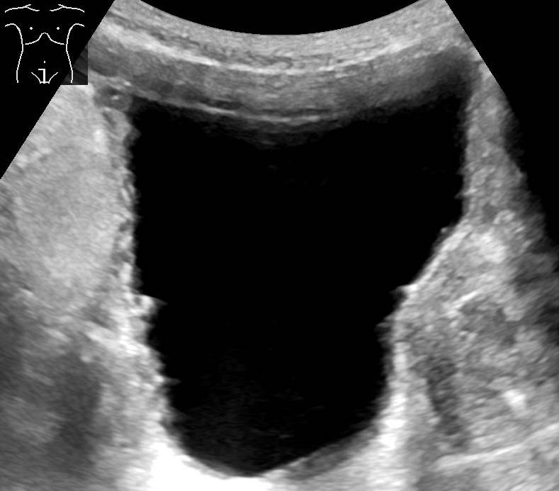
A full bladder on a suprapubic scan. The urine reads black and presents the wall for inspection. This wall has thickened into ridges from long strain against an outlet block.
A bladder tumor is a growth on the inner wall of the bladder. Ultrasound shows it as a solid lump pushing into the urine. The urine sits black on the screen. A tumor stands out as a pale shape rooted to the wall. A handheld scanner finds that shape at the bedside in a few minutes. The probe sits low on the belly, above the pubic bone, and looks down into a full bladder. The bladder makes its own window. A bag of urine carries the sound to the far wall and lights up anything growing from it. A tumor shows as a knot of echo on a wall that should look smooth. Many growths rise from the lining as a delicate frond on a stalk. The frond sways a little in the urine under the probe. Other growths sit as a broad mound on a wide base. A broad mound carries a higher chance of growing deep into the wall. The scan catches the larger growths of either kind plainly. A clinician measures a mass in two directions and counts how many sit on the wall. The number and the size go into the note. The finding then sends the person for a cystoscopy and a closer look.
Blood in the urine brings people to this scan. A painless show of visible blood is the common first sign of a bladder tumor. The urine runs red for a day. It clears the day after. The blood returns weeks later. The calm of it fools people into waiting. A clinician treats any visible blood in the urine of an adult as a reason to look inside the bladder. The scan starts that search at the bedside. It checks the bladder wall for a mass before the person waits weeks for a clinic slot. Blood too faint to see can still show on a dipstick and bring the same need to look. The flat tumors can bring a burning urgency of their own. A single episode of visible blood counts on its own, even after the urine clears. Visible blood in the urine carries a cancer in about one person in eight. The odds climb in older people, in smokers, and in workers exposed to dyes and rubber. The odds climb again in regions where a bladder fluke lives in the water. The scan turns a vague worry into a finding a clinician can act on.
A bladder mass shows the same on a handheld and on a cart machine. A large frond stands out plainly on a coarse picture. A clinician new to bladder scanning learns the look early. The bladder lies shallow below the navel. A probe reaches it with ease. A full bladder brings the whole wall into view. The scan finds a mass, measures it across, and notes where it sits on the wall. A clinician records the size, the site, and the number of masses in the note. A clinician also marks any mass that sits near a ureter opening. A clinician saves a still image of the mass for the referral and for the next visit. A picture in the note carries more than a line of text. A nurse, a junior doctor, or a clinic far from a hospital can each run this scan. The bladder gives up its secret to a simple look. The work of naming, grading, and staging waits for the specialist.
Filling the bladder for the look
The scan needs a full bladder. A full bladder spreads the wall thin and lifts it into view. A thin wall reveals a small mass. A clinician asks the person to drink and hold, or fills the bladder through a catheter when speed matters. Thick folds in a half-empty bladder can bury a small tumor. A clinician fills the bladder first and scans on the full state. The probe sits above the pubic bone and aims down into the pelvis. A clinician sweeps the bladder from side to side in one plane. The clinician turns the probe ninety degrees and sweeps top to bottom in the other plane. Two planes cover the whole wall. A clinician notes the bladder volume from the same full scan. The urologist uses that figure later. A mass on the front wall, the back wall, the dome, or the floor each comes into a clear view this way. A clinician lingers on any spot that looks raised and studies it from two angles. The scan covers both walls and the roof in a minute.
Some parts of the bladder hide a tumor better than others. The dome at the top sits close to the probe and can fall in a near-field blur. The front wall behind the pubic bone sits under reverberation lines. A clinician tilts the probe and changes the fill to bring these zones out. The trigone at the floor holds many tumors and deserves a careful sweep. A clinician angles down toward the trigone and watches the two ureter openings. A jet of urine spurts from each opening on a working side. A small tumor near a ureter can block it and swell the kidney above. The scan checks both kidneys on the same visit for that swelling. A swollen kidney over a bladder mass adds weight to the urgency. A heavy build pushes the bladder deep and dims the picture. A clinician drops the frequency and presses in to bring the wall back. A careful operator sweeps every wall before calling a bladder clear.
Tumor, clot, or stone
Three things make a lump in the bladder. A tumor grows from the wall. A blood clot floats free. A stone rolls on the floor. Ultrasound separates them with two simple tests. The first test moves the patient. The second test adds color Doppler. A clinician rolls the person from flat onto one side and watches the lump. A tumor stays fixed to the wall through the roll. The scan shows it as part of the wall. The base holds it in place. A clinician then opens color Doppler over the lump. A tumor lights up with its own blood supply. A stalk of vessels runs into its base and feeds it. A papillary frond shows a single vessel climbing its stem. The flow runs strongest at the base where the stalk enters. A larger tumor pulls a richer tangle of vessels into itself. A fixed base and a living blood supply together mark a growth that needs a cystoscopy.
A blood clot makes a lump that looks solid at first. A clot forms a loose clump of echo. It has no feeding stalk. It throws no shadow. A clinician rolls the person and watches the clot slide to the low side of the bladder. A clot moves with gravity and settles where the floor now lies. Color Doppler over a clot stays dark. The clot holds no vessels of its own. A clinician can flush the bladder through a catheter and watch a clot break up and shift. A clot also changes from one scan to the next. It forms and dissolves over hours. A clot left from an old bleed can shrink and darken over days. A fresh bleed can fill the bladder with floating debris and hide the wall behind it. A clinician clears that blood and scans again on clear urine. A free, dark, drifting clump points to blood. A clinician calls it a clot and clears it with a flush.
A stone in the bladder makes a bright lump with a dark shadow behind it. The shadow is the giveaway. Sound bounces off the hard stone and leaves a black stripe below. A clinician rolls the person and watches the stone tumble to the low point. A stone moves like a clot and shadows like bone. Color Doppler behind a stone can throw a flicker of color on some machines. The rough surface makes that twinkle. A stone forms in urine that sits too long behind a block. A stone sits loose in the urine and answers to gravity every time. A large stone can scrape the wall and bring its own blood into the urine. A clinician notes the stone and still checks the wall around it. A bright lump with a tail of shadow rolls free in the urine. A clinician calls it a stone.
The three lumps sort out fast at the bedside. A clinician moves the patient, opens Doppler, and looks for a shadow. A fixed lump with its own blood flow points to a tumor. A drifting dark clump points to a clot. A bright lump with a shadow points to a stone. A clot can hide a tumor underneath it on a bladder full of blood. A clinician clears the blood and scans again before calling the wall clean. A tumor that lies flat can defeat the rolling test. It barely lifts off the wall. A clot stuck to the wall by fresh fibrin can sit still for a time. A clinician leans on Doppler when movement alone leaves a doubt. A second scan after a bladder flush clears the doubt in many cases. A clinician sends any lump that stays unclear on for a cystoscopy. The camera settles what the scan leaves open. A confident call at the bedside spares a person a needless wait.
A mass that does not move
One test sorts the three lumps. A clinician rolls the patient and watches the wall. A tumor stays anchored where it grows.
Reading the wall and the base
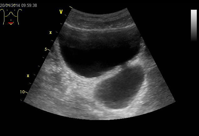
A bladder diverticulum on a transverse scan. The large black space is the full bladder. The rounded pouch below it is a diverticulum, an outpouching that can mimic a mass beside the bladder. The marks down the left edge are the depth scale in centimeters.
The bladder wall carries its own signs. A normal wall shows as a smooth line under three millimeters on a full bladder. A clinician measures the wall on a bladder at least half full. A poorly filled bladder thickens its wall on its own and shows a falsely high figure. A clinician fills the bladder and measures again before trusting a thick wall. A wall thickened all over points to a bladder strained behind an outlet block for a long time. A focal lump that breaks the smooth line of the wall raises the question of a tumor. A clinician measures a focal thickening and watches it over time. A growing patch of wall earns the camera. In regions where the bladder fluke lives in the water, the parasite scars the wall, thickens it, and lays down a rim of calcium. A bladder marked by that fluke can carry both calcium and a raised cancer at once. A bright line of calcium with a focal mass deserves a close look and a referral. A clinician sends a focal thickening on for a cystoscopy.
Some lumps at the base are not tumors at all. The prostate can bulge up into the bladder floor in an older man. This median lobe rises as a round mound at the midline. A layer of intact wall covers it. A clinician judges its shape and its place at the neck and calls it prostate. A pouch in the wall, a diverticulum, can hold debris that mimics a mass. A tube of swollen ureter, a ureterocele, can balloon into the floor near its opening. An inflamed bladder thickens its whole wall and roughens the lining on its own. A recent infection explains that change. A folded wall in a half-empty bladder can throw up a false lump that melts away on filling. A clinician judges each by its shape, its place, and its link to the structures around it. A benign lump carries a known shape and a known home. A clinician saves the close worry for a mass that grows from the lining itself.
Bladder wall and mass: numbers at the bedside
What is measured
Value
What it points to
Bladder wall, well filled
under 3 mm
normal
Bladder wall, near empty
up to 5 mm
refill, then re-read
Fill before reading the wall
half full or more
a true measurement
Ultrasound in visible hematuria
87 to 93% sensitive
reliable for raised tumors
Tumor under 5 mm or flat (CIS)
often missed
cystoscopy still needed
Visible painless blood in urine
a cancer in about 1 in 8
refer for cystoscopy
What the scan cannot settle
The scan finds a mass. It cannot name what the mass is made of. A bladder tumor ranges from a low-grade growth that rarely spreads to a high-grade cancer that invades the muscle. The two look alike on the scan. The biggest question for a tumor is its depth. A growth held to the surface carries a far lighter outlook than one grown into the muscle. A clinician takes the size, the site, and the number from the scan. The grade and the depth wait for tissue under a microscope. A handheld can hint at deep growth when a mass breaks through the wall layers. The proof of that depth waits for the camera and the lab. Only a cystoscopy and a biopsy name the tumor for certain. A urologist passes a camera into the bladder, sees the mass directly, and takes a piece for the lab. The lab names the cell type and the grade. A clinician treats the scan as the door to that path. The path runs on to the urologist and the lab.
The scan misses some tumors. A small tumor under half a centimeter can hide in a fold or a blind spot. A flat tumor that spreads along the lining, a carcinoma in situ, leaves no lump to catch. Ultrasound shows the wall as normal over a flat cancer. The scan catches the raised tumors of a centimeter or more far better. Studies of people with visible blood in the urine put ultrasound around nine in ten for a tumor. The miss falls on the small and the flat. A urine test for tumor cells runs alongside the scan and helps with the flat ones. A clear scan does not rule out a tumor on its own. A clinician still sends a person with visible blood in the urine for a cystoscopy after a clear scan. A clinician also orders a CT scan of the upper tracts, since a tumor can grow in a kidney or a ureter beyond the reach of the bladder probe.
The scan sits inside a larger workup. A person with visible blood in the urine needs a full look at the whole urinary tract. The bedside scan starts that look in minutes. It checks the bladder wall for a mass. It checks both kidneys for a backup. It checks the volume of urine left after the person voids. The scan hands the clinician a first map of the problem. A mass on the wall sends the person to a urologist on an urgent line. The urologist books a cystoscopy to see the mass directly. The cystoscopy shows the surface, the base, and the spread of the tumor across the lining. A urologist takes the tumor out through the camera in a procedure called a TURBT. The TURBT removes the growth and supplies the tissue for the lab in one step. The lab names the cell type, the grade, and the reach of the tumor into the wall layers. Bladder cancer often grows in more than one place at once. A urologist surveys the whole lining at the camera for a second tumor and a third. A CT scan of the abdomen and the pelvis maps the kidneys, the ureters, and any spread beyond the bladder. The CT runs a dye through the urinary tract and catches a second tumor higher up in a kidney or a ureter. The team builds a stage and a grade from the camera, the lab, and the CT together. A clinician explains the plan once those pieces land. Treatment follows the grade and the depth, from a TURBT and a watch at the light end to medicine into the bladder or a major operation at the heavy end. The deepest tumors call for removing the bladder itself. The handheld scan does none of this naming and staging. It opens the door and sets the speed. A mass found at the bedside today moves the person onto the urgent path today.
Bladder tumors come back often. A person treated for a bladder tumor needs a watch for years. The same lining that grew the first tumor can grow a second. A urologist sets a schedule of cystoscopies to catch a new tumor early. The handheld scan fits between those camera checks. A clinician scans the wall for a new lump at a routine visit. A clinician tracks the size of a known mass across visits and flags any growth. A scan that finds a recurrence sends the person back to the camera ahead of schedule. The bedside check adds a set of eyes between the formal looks. A missed recurrence grows in the gap between two camera dates. A clear handheld scan still leaves the cystoscopy in place on its schedule. The two work together over the long watch a bladder cancer needs.
The scan knows its place in all this. It finds a mass and raises the alarm. It cannot grade the tumor or stage it. It cannot stand in for the camera or the biopsy. A clinician takes the scan for what it gives. It gives an early warning from the bedside. A clinician hands that warning to the urologist with the size, the site, and a picture. The rest of the answer comes from the urologist and the lab.
The handheld at the bedside
The handheld brings this scan to the person. A clinic with no machine of its own checks a bladder at the bedside. A nurse scans a person with blood in the urine on the ward. A rural clinician checks a bladder wall where the nearest cystoscopy sits a day’s travel away. The scan runs in minutes on a wand the size of a phone probe. It needs only a full bladder and a quiet corner. A clinician finds a mass, measures it, and sends the person on the right path. A bladder cancer caught at a first bleed carries the best odds of a cure. A handheld scan at a first visit can shave weeks off the road to that diagnosis. The handheld puts that first catch within reach of more people.
The scan costs little to repeat. A clinician scans a bladder today and again next month at no risk to the person. A bladder under a watch shows a new lump on a routine visit. A person on treatment shows the bladder calming or the tumor returning over a run of scans. The same wand that found the first tumor follows the bladder for years. A handheld carries that long watch out of the hospital and into the clinic. The check travels to the person each time. A short clip stored from each visit lets a clinician compare the wall month to month.
The handheld does one clear job here. It finds a mass on the bladder wall and starts the clock. A clinician takes the size, the site, and the movement, opens color Doppler, and decides who needs the camera next. The scan hands the person a head start on a bladder cancer. The urologist and the lab carry the diagnosis from there. The bedside look gets the right person to that door, in the few minutes a handheld takes.
Common questions
Can ultrasound find a bladder tumor?
Yes, ultrasound finds raised bladder tumors of a centimeter or more. A full bladder shows the wall. A tumor appears as a solid lump growing from it. In people with visible blood in the urine, ultrasound catches a tumor around nine times in ten. Small tumors under half a centimeter and flat tumors slip past. A clear scan still needs a cystoscopy to confirm.
How does ultrasound tell a bladder tumor from a blood clot?
A clinician uses two simple tests. The first is movement. The second is color Doppler. A tumor stays anchored to the wall through a change of position. A tumor carries its own blood supply and lights up on Doppler. Those two signs together mark a growth. A blood clot gives a different picture. A clot slides with gravity to the low point of the bladder. A clot stays dark on Doppler. It holds no vessels of its own. The anchored lump with its own flow is the tumor. It earns a cystoscopy.
Why does the bladder need to be full for the scan?
A full bladder makes the window for the scan. Urine carries the sound to the far wall. A filled wall spreads thin and shows a small mass. A poorly filled bladder folds its wall into thick pleats. The pleats hide a small tumor and show a falsely thick wall. A clinician fills the bladder before measuring the wall.
Does a normal bladder scan rule out cancer?
No. Ultrasound misses small tumors under half a centimeter and flat tumors that spread along the lining. A clear scan lowers the worry only so far. A person with visible blood in the urine still needs a cystoscopy after a normal scan. The camera sees the flat and the small lesions directly. A clinician treats a clear handheld scan as a start. The cystoscopy gives the all-clear.
About the Author
Julien Mercier
Senior R&D Engineer · Medical Ultrasound Transducer Development
Senior R&D Engineer with an M.S. in Applied Physics and over 15 years of experience in medical ultrasound transducer development, specializing in the design verification and performance testing of high-frequency imaging transducers. Currently leading the development and verification of the company’s next-generation high-frequency linear-array transducer, responsible for imaging performance evaluation and reliability analysis in preclinical testing. Brings extensive hands-on experience in piezoelectric element tuning, beamforming parameter optimization, and system-level performance testing.