Prostate volume is the size of the gland in cubic centimeters, worked out on the ultrasound scan from three measurements of the prostate. The number matters in its own right. A larger gland makes more PSA across its bulk, so the volume helps read a raised blood test. It also guides the treatment of an enlarged prostate. Measuring it is a quick step in a routine transrectal scan.
Why volume is measured
Prostate volume earns its place through the PSA. A raised PSA can come from cancer. It can also come from a gland that has grown large. A big prostate makes more PSA across its bulk. The volume lets a doctor read a PSA against the size of the gland behind it. A PSA reading alone leaves out the size of the gland. The volume supplies that size. The two read together give the number its meaning.
The volume turns a bare PSA into a sharper figure. Dividing the PSA by the volume gives the PSA density. A high PSA from a large gland reads as less worrying once the size is known. The volume is the number that unlocks that reading. It is the scan’s main numerical contribution to the workup. Without the size, the PSA hangs in the air. The volume anchors it to the gland. One quick measurement does that work.
Volume matters for the enlarged gland too. An aging prostate grows, pressing on the urethra and slowing the urinary stream. The size guides the choice of medicine or surgery for that enlargement. A surgeon plans the operation around the gland’s volume. The same measurement serves the cancer workup and the enlargement. An older man often has both questions at once. The one volume reading answers each of them. The measurement does double duty in a single scan.
The ellipsoid formula
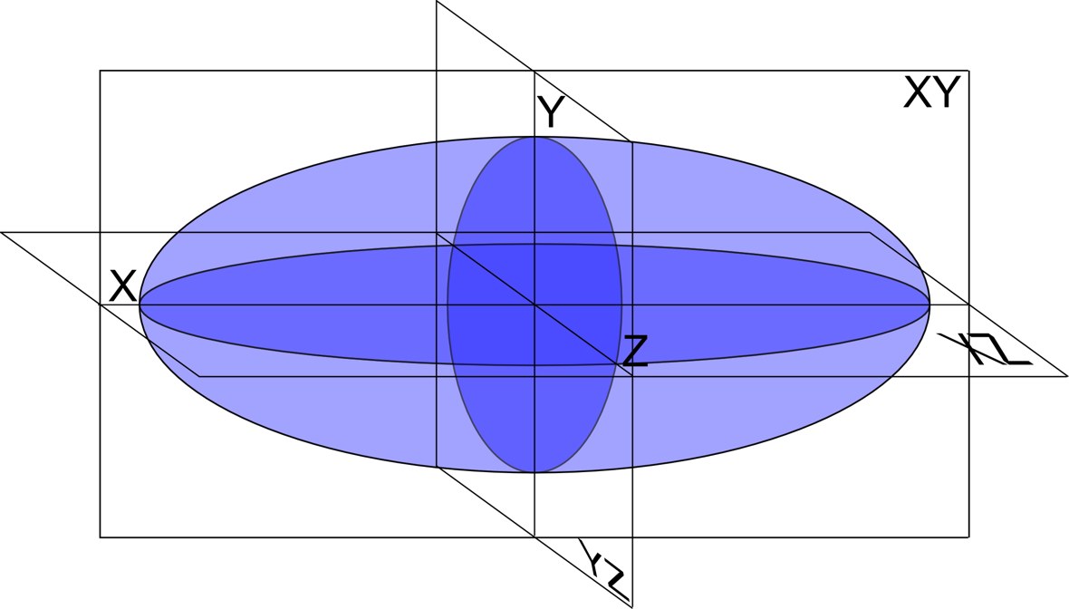
The volume is worked out with a simple formula. The prostate is treated as an ellipsoid, a rounded egg shape. Its volume is its length times its width times its height, multiplied by a fixed factor of 0.52. Three quick measurements feed the sum. The scanner often does the arithmetic once the diameters are entered. The sum is plain arithmetic. Three lengths multiplied, then scaled by a constant, give the volume. A pocket calculator could do it.
The formula is the standard across ultrasound. A urologist measures the three diameters and reads the volume off the calculation. The 0.52 factor comes from the geometry of an ellipsoid. The method is fast enough to do at every scan. It gives a usable volume in under a minute. The speed is part of why the formula endures. It fits into any scan, busy list or not. A method that took ten minutes would never have lasted.
Working out a prostate volume: a worked example
Measurement
Example value
Length (base to apex)
4.5 cm
Width (side to side)
4.0 cm
Height (front to back)
3.0 cm
Formula
length × width × height × 0.52
Volume
about 28 cubic centimeters
PSA density (for a PSA of 4.0)
4.0 ÷ 28 ≈ 0.14
Measuring the three diameters
The prostate is measured as an ellipsoid, a rounded egg shape with three axes. Its length, width, and height are the diameters the volume formula multiplies together with the factor 0.52.
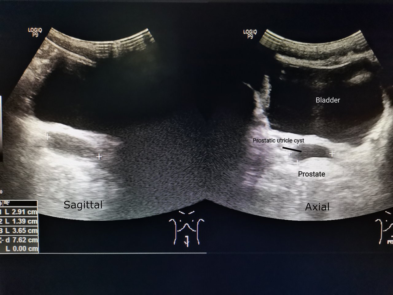
The three measurements come from two views of the gland. The probe shows the prostate in a side-on sagittal plane and in a cross-wise axial plane. The length runs from the base down to the apex. The width runs from one side to the other. The height runs from front to back. The three directions sit at right angles to each other. Each is the longest distance across the gland in its plane. Together they frame the gland as a box around the egg shape.
Each diameter is read with on-screen calipers. The urologist freezes the picture and drops a caliper at each end of the measurement. The screen reads off the distance in centimeters. The same is done for all three diameters. Care over the caliper placement is what makes the volume reliable. A caliper dropped a few millimeters off shifts the result. The error in each diameter feeds into the whole sum. Steady placement on a frozen frame keeps it honest.
The length and the height come from the sagittal view. A clean mid-line sagittal picture shows the gland from base to apex and from front to back. The urologist measures both diameters on that one frozen frame. A good sagittal plane repays the time taken over it. Two of the three numbers ride on it. A urologist takes care to find the true mid-line. An off-centre plane reads the gland short. The mid-line sagittal frame is the one to get right.
The width comes from the axial view. The probe turns to show the gland in cross-section at its widest. The urologist measures side to side across that view. The widest slice gives the true width. The three diameters together feed the volume. The axial width is read at the broadest cut of the gland. A slice taken too high or too low reads narrow. The widest cross-section gives the figure the formula wants.
PSA density, the main payoff
A prostate on transrectal ultrasound, shown in the sagittal and axial planes the three diameters are read from. The bright marker is an incidental benign cyst.
PSA density is the main reason the volume is measured. It is the PSA divided by the volume of the gland. The figure gives the amount of PSA for each cubic centimeter of prostate. It reads a raised PSA in the light of the gland that made it. The density sharpens a borderline blood test. It reads a PSA in proportion to the gland that made it. The size that the volume supplies is what makes that proportion possible.
The density puts a PSA in proportion to the gland. A lot of PSA coming from a small gland gives a high density. That high density is the figure that worries a doctor. It points toward a significant cancer behind the reading. The volume is what lets a doctor read a PSA in proportion this way. The number carries a meaning the bare PSA cannot.
A density threshold guides the biopsy decision. A widely-used threshold sits near 0.15 nanograms per milliliter for each cubic centimeter, below which significant cancer is less likely. A reading above it pushes a man toward a biopsy. The figure feeds into whether a biopsy is needed. The volume from the scan is what makes the density possible. The threshold is a guide read alongside the rest of the picture. A density near it leaves room for judgment. The figure informs the decision a doctor still makes.
PSA density saves some men a biopsy. A reassuring density, read with the rest of the picture, can support watchful testing. The figure helps spare men a procedure they may not need. The volume measurement carries real weight in that decision. The caliper measurement is quick, and the number it feeds carries real weight. An avoided biopsy is a real benefit of the density. A man kept off the table is spared its risks. The volume behind that density did real work.
PSA density also tracks a gland over time. A man on active surveillance has his density checked at intervals. A rising density can signal a change that earns a closer look. The volume is remeasured each time to keep the density current. The measurement stays useful long after the first scan. The trend in the density matters more than any one reading. A stable density reassures over the years. The remeasured volume keeps that trend honest.
The 0.52 factor and its shape
The 0.52 factor comes from treating the gland as an ellipsoid. The volume of an ellipsoid is its three diameters multiplied together and by a constant. That constant works out to about 0.52. The formula assumes the prostate is a smooth egg shape. Many glands are close enough to that shape for the formula to serve. The constant is fixed for every gland. It holds the same for every prostate. The same 0.52 turns three diameters into a volume each time.
The shape is a useful approximation of a real gland. A real prostate is lumpier than a smooth ellipsoid. The factor suits the average gland. A much-enlarged gland strays further from the ellipsoid shape. The formula carries a known margin of error for that reason. The error is the price of the formula’s speed. A doctor accepts it for a number in under a minute. The approximation is good enough for the uses it serves.
A walnut, an orange
A volume reading is easier to picture in everyday terms. A young man’s prostate is about the size of a walnut, near twenty cubic centimeters. An enlarged gland can swell to the size of an orange or larger, past eighty. The number on the screen places a gland on that scale. A doctor reads the volume to know whether a gland sits in the normal range or well beyond it. The everyday comparison helps a man picture his own result. A volume near twenty takes on a real size once it is named a walnut. The image gives the number a size a man can feel.
Guiding treatment of an enlarged gland
Volume steers the treatment of an enlarged prostate. The medicines for a large gland are chosen partly by its size. A drug that shrinks the gland suits a man whose prostate is clearly enlarged. The volume tells the doctor how big the problem is. The number guides the first treatment choice. The size of the gland sets the first step. The volume tells the doctor which one a man has. The size shapes the medicine from the start. The volume places a man on a clear scale before treatment is chosen. A gland past eighty cubic centimeters is plainly enlarged. The size names the problem in a number the doctor acts on.
Surgery for enlargement leans on the volume too. The operation to relieve a blocked stream is planned around the gland’s size. A greatly enlarged gland may call for a different approach from a modest one. The surgeon reads the volume before choosing the method. The measurement shapes the plan for the gland. A surgeon needs the size before picking the operation. The volume sets expectations for the procedure. The number is part of the plan a man is given.
Accuracy and the sources of error
The volume from the formula is a close estimate. The ellipsoid shape only approximates a real gland. The caliper placement adds its own small error. The result sits close to the truth. A doctor reads the volume as a good working figure. No bedside method gives a perfect volume. The formula trades a little accuracy for a lot of speed. A doctor reads the result in that light.
The ellipsoid formula tends to under-read the gland. According to a study comparing the formula with planimetry, the ellipsoid method underestimates true prostate volume by around eighteen percent. The under-read is larger for a bigger gland. A doctor allows for that bias in reading the figure. The number runs a little low. The bias is steady and its direction is known. A steady bias is easier to live with than a random one. The doctor knows the figure runs low and reads it so. A predictable error is a manageable one.
Good technique keeps the error small. A clean sagittal plane and a proper axial view at the widest point, with careful calipers on each, give true diameters. The volume is only as good as the three measurements behind it. A sloppy plane feeds a poor number. The hard part is the planes. The machine does the sum. The urologist’s job is the clean views and the careful calipers.
The known bias does not undo the volume’s value. A figure that runs low in a steady way is still useful for PSA density and for tracking. A doctor reads it knowing its habits. The estimate serves the decisions it feeds. A consistent measurement matters more than a perfect one. What the volume feeds needs consistency above all. A density tracked over years leans on a steady method. The formula gives that steadiness scan after scan.
Other ways to measure
The ellipsoid formula has alternatives. A planimetry method traces the gland on many slices and adds them up. It traces the gland slice by slice for a truer volume. The cost is more time on each scan. The bullet-shape formula uses a different factor for a closer fit. Each method trades speed for accuracy in its own way. The everyday scan rarely needs the slower methods. The ellipsoid figure is close enough for the density and the tracking. The fuller methods come out for special questions.
Three-dimensional and automated tools have grown more common. A 3D probe can sweep the gland and compute a volume directly. Software can trace the outline and do the arithmetic. These methods cut the hand measurement out of the estimate. The ellipsoid formula stays the everyday standard for its speed. The newer tools promise less hand work. A swept volume removes the caliper error. The simple formula holds its place by being the quickest method to hand.
Volume on a handheld probe
A handheld ultrasound measures volume as readily as any machine. A transrectal probe run from a tablet shows the two planes and the calipers. The urologist measures the three diameters on the screen. The tablet computes the volume from the formula. The whole measurement takes a minute at the couch. The handheld machine carries the same formula. The calipers and the calculation are built into the app. A urologist measures the gland with the scanner in one hand.
The handheld setup folds the volume into the visit. A man having a prostate scan gets his volume measured in the same sitting. The PSA density can be worked out there and then. A urologist reads the size with the gland on the screen. The measurement adds little time to the scan. The volume comes alongside the scan that reads the gland. A urologist already in the prostate takes the three measurements in passing. The figure is ready before the probe comes out.
Prostate volume is one small measurement that many readings lean on. It feeds the PSA density, it sizes the gland for treatment, and it tracks a prostate over time. A handheld probe takes it in a minute. The quick set of calipers travels to wherever a man is scanned. A small clinic measures volume as well as a large one. The formula needs no special hardware. A probe and a tablet are enough.
The volume in the whole assessment
The volume sits quietly inside the prostate workup. It rarely makes a diagnosis on its own. It sharpens the PSA, sizes the gland, and feeds the plan. The number works in the background of the larger picture. Its value lies in what it enables. The volume is a means to other readings. It serves the density, the treatment plan, and the tracking. The figure matters through the decisions it supports.
A reliable volume repays the care it takes. A measurement done carefully feeds a PSA density a doctor can trust. The minute spent over the calipers pays off down the line. The figure carries into every reading that uses it. The volume is a small craft done well. The care shows up later, in numbers a doctor can rely on. A density, a treatment choice, and a trend all rest on the volume. A minute of care holds all three up.
Prostate volume, in the end, is three quick measurements and a simple sum. From them comes a number that reads a PSA, sizes a gland, and tracks a change. On a handheld probe at the bedside, that small measurement is always at hand. The volume is the quiet figure much of the prostate workup leans on. From three lengths and a constant comes a figure with real reach. It reads a blood test, sizes a gland, and follows a change over years. The handheld probe keeps that figure within reach at any couch.
Common questions about prostate volume
How is prostate volume measured on ultrasound?
From three diameters of the gland, read with on-screen calipers. The length and height come from a side-on sagittal view, the width from a cross-wise axial view. The three are multiplied together and by a factor of 0.52, the ellipsoid formula. The scanner usually does the arithmetic once the measurements are entered.
What is the formula for prostate volume?
Length times width times height times 0.52. The 0.52 comes from treating the gland as an ellipsoid, a smooth egg shape. A gland measuring 4.5 by 4.0 by 3.0 centimeters works out to about 28 cubic centimeters. The same formula serves on any ultrasound machine.
Why does prostate volume matter?
It unlocks the PSA density, the PSA divided by the volume, which reads a raised blood test against the size of the gland. A high PSA from a large gland is less worrying once the size is known. The volume also sizes an enlarged gland for the choice of medicine or surgery. One quick measurement feeds both.
Is the ellipsoid formula accurate?
It gives a close estimate. The figure runs a little low, in a steady and known way. Studies comparing it with slice-by-slice planimetry find it underestimates true volume by around eighteen percent, more so for a large gland. The bias is steady and in a known direction, so the figure stays useful for PSA density and for tracking. A consistent measurement matters more than a perfect one.
What is a normal prostate volume?
A young man’s prostate is about the size of a walnut, near twenty cubic centimeters. The gland tends to grow larger in older men. An enlarged prostate can reach the size of an orange or larger, past eighty cubic centimeters. The volume places a particular gland on that scale.
About the Author
Julien Mercier
Senior R&D Engineer · Medical Ultrasound Transducer Development
Senior R&D Engineer with an M.S. in Applied Physics and over 15 years of experience in medical ultrasound transducer development, specializing in the design verification and performance testing of high-frequency imaging transducers. Currently leading the development and verification of the company’s next-generation high-frequency linear-array transducer, responsible for imaging performance evaluation and reliability analysis in preclinical testing. Brings extensive hands-on experience in piezoelectric element tuning, beamforming parameter optimization, and system-level performance testing.