Skin layer ultrasound analysis is the use of a high-frequency probe to measure the layers of the skin, the epidermis and the dermis. The probe reads each layer as a separate band on the screen. It puts a number on the thickness of each and on the texture of the dermis. The skin runs only a few millimeters deep. These figures turn what a clinician could once only look at and feel into something measured, recorded and followed across the months and years.
Higher frequency, finer skin
The reading lives or dies on the frequency of the probe. Splitting the skin into its layers takes a high frequency. A probe at 18 to 24 MHz resolves a structure well under a millimeter, fine enough to part the epidermis from the dermis and to measure the thickness of each. The detail falls away at the lower frequencies a body probe uses, where the skin reads as one bright line on the fat. The skin sits in the first few millimeters under the surface, the exact reach where this frequency does its best work. Frequencies higher still, used on the surface in research and in some clinics, sharpen the epidermis further. The handheld probe a clinic carries works in the band where the whole skin comes into one picture. Frequency comes at a cost. The higher it climbs, the shallower it reaches, since high-frequency sound fades fast in tissue. A probe tuned to read the skin in fine detail gives up the depth a body probe has. That trade suits the skin, which sits in the first centimeter and asks for detail over reach. A clinic that wants the deeper tissue as well keeps a second probe, each one set for the depth its target sits at.
The dermis is where the story is
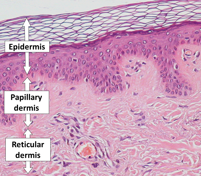
The skin under the microscope: the epidermis on top, the papillary and reticular dermis below. The dermis is the broad layer that lights up on ultrasound, where its collagen reflects the sound. This is a histology slide, not an ultrasound image.
The skin reads as a stack of bands. The dermis is the band that matters here. It sits below the bright top line of the epidermis and reads as the broadest layer on the screen. It is also the brightest. The brightness comes from collagen. The packed fibers of the dermis throw the sound back strongly, so a dermis full of well-ordered collagen lights up. That brightness is the dermis reporting on its own substance. The more collagen it holds, the brighter it reads. The layer carries the strength of the skin, the spring in it, and the bulk a treatment or a disease changes. Collagen brightness is the heart of the reading. A young dermis packs its collagen tightly, in ordered bundles that send a strong echo back. Age, sun and disease break that order down. The echo weakens with it. The scan reads the brightness as a stand-in for collagen a clinician cannot see directly. Lost collagen shows as a dimmer dermis, a layer giving up some of what holds the skin’s strength.
Above the dermis runs the epidermis, the thin outer layer. On the screen it is the entry echo, the first bright line where the sound meets the surface. Over much of the body the epidermis is a fraction of a millimeter, too thin to read as more than that line. It grows thicker on the palm and the sole, thick enough there to measure on its own. The line marking the top of the skin is sharp on a clean scan. The dermis begins just below it. Reading where one ends and the other starts is the first thing the probe does that a look at the surface cannot. Where the whole skin is shallow, as on the eyelid, the layers crowd together. Only a high frequency keeps them apart. The probe reads the epidermis where there is enough of it to read, and notes the entry line where there is not.
Under the dermis the brightness falls away. The fibers thin out, the collagen gives way, and the layer darkens into the fat below. That drop marks the floor of the skin. The fat beneath it has its own thickness and its own uses, measured in the body work a clinic does for contour. This reading stops at the floor. It stays in the skin proper, the epidermis and the dermis, where aging and disease and scars write themselves. The fat below is a separate layer with a separate story. Keeping the two apart matters for the reading. A measurement that wanders into the fat reports a thickness the skin does not have. The clinician sets the calipers from the entry line down to the floor of the dermis, and stops there. The fat is read on its own terms when a clinic wants it, with the gain and the depth set for a layer that sits deeper and reads darker.
The whole skin is a few millimeters deep. The probe reads it from the surface down, layer by layer.
Two numbers: thickness and brightness
Thickness is the first figure. The probe reads how deep the dermis runs and how thick the epidermis sits over it. Both vary by site. Eyelid skin is thin. The skin of the back is thick. Both shift with age and with the sun a person has carried over a lifetime. A thickness measured today gives a baseline, a fixed number that a later scan can be set against at the same spot. A first thickness is only a starting mark. Its value comes when the same spot is measured again and the two figures sit side by side, the change between them read off at a glance.
Brightness is the second figure. It is the more telling of the two. The brightness comes from collagen: the more the dermis holds, the brighter it reads, and the less it holds, the darker. The scan can put a value on that brightness, counting how much of the dermis falls below a chosen level of echo. A rising count of dark pixels in the dermis is one of the clearest signals the scan follows, the dermis losing its substance in a number. The count goes by names like the number of low-echogenic pixels, the share of the dermis reading below a set brightness. The more collagen is lost, the higher the count climbs, which gives a clinician a single value to follow from one visit to the next. The count puts a firm number on a dimming the eye reads only as vague. That figure is what carries the reading from a guess to a measurement a clinic can act on.
The pattern is read alongside the two figures. A settled dermis shows an even band. Disease, fluid or a scar breaks that evenness up. The probe shows where a darker zone has crept in. The eye reads the pattern. The machine reads the figures. Together they describe a state of the skin that no single look or pinch arrives at.
The sun writes a band no mirror shows
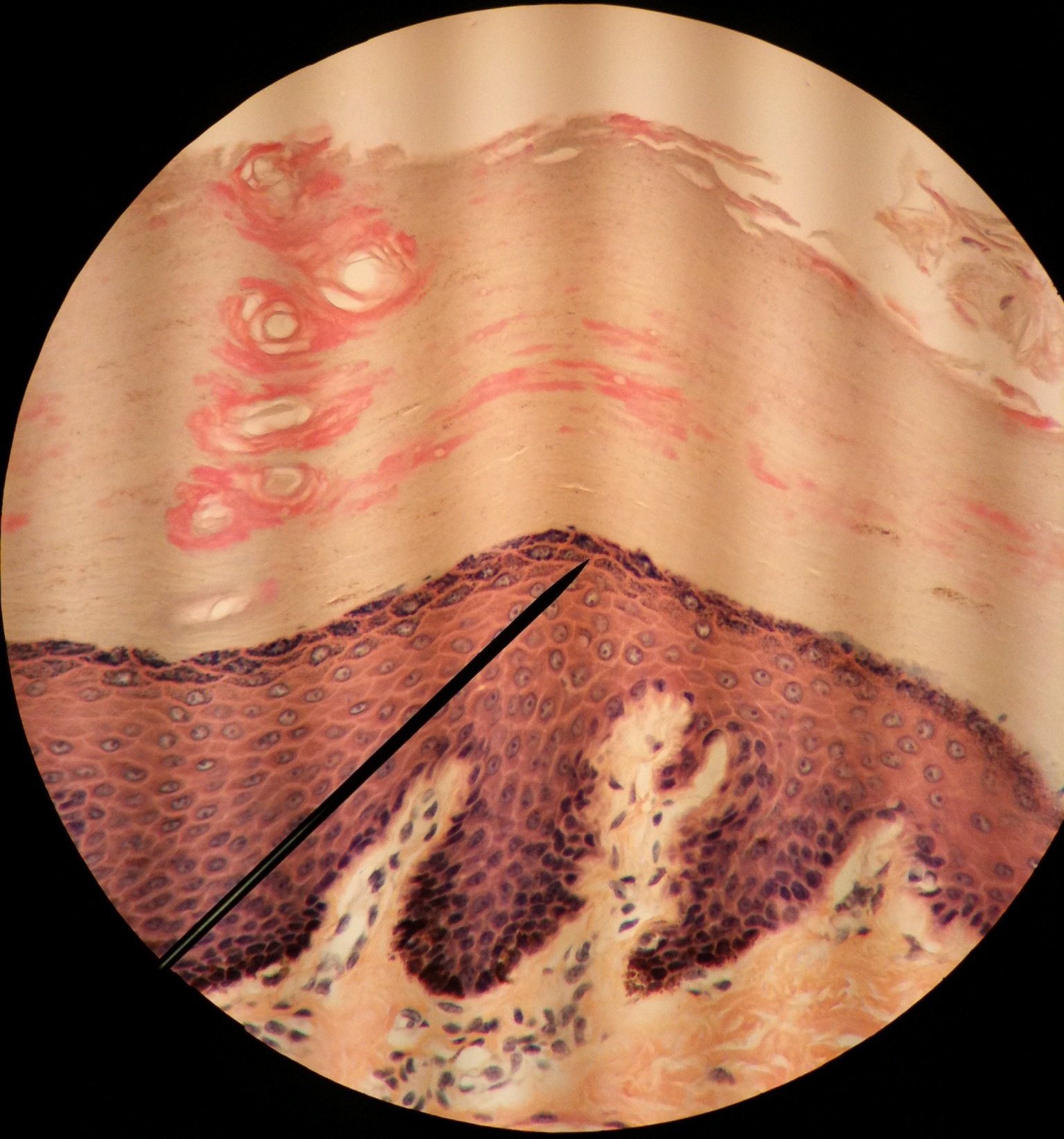
Thick skin from a palm or a sole, its heavy keratin layer above the epidermis and the dermis. A high-frequency probe reads these same layers as bright bands without a biopsy. This is a histology slide; the dark diagonal is the microscope’s pointer.
Years of sun leave a mark on the skin. The scan reads it better than the eye can. A dark band gathers just under the epidermis, lying on top of the bright dermis. Dermatology calls it the subepidermal low echogenic band. It builds in skin that has taken heavy sun across a lifetime, from damaged fibers and the material that collects with photoaging. The more sun a skin has taken over the years, the broader the band. On skin kept covered it stays faint. The band is more than a marker of age. It is a record of exposure. Two patients of the same age show different bands if one lived in the sun and the other stayed out of it. The width of the band reads that history off the skin. A clinician can point to it on the screen when a patient asks why one part of the face has aged faster than another, the answer often lying in the years of light that part has taken.
The dermis ages underneath it. The collagen that gave the band its brightness is lost, so an older sun-worn dermis reads thinner and darker than a young one. The two readings move together, the dark upper band widening as the bright dermis fades. The scan puts both into numbers. The numbers track how far the skin has aged past what its surface lets on. The pairing is what makes the reading strong. A dark band alone could be many things. Sitting over a dermis thinned and dimmed, it reads clearly as photoaging, the two findings backing each other. The scan reports them together. The picture of sun-aged skin is firmer for it.
This is the reading an aesthetic clinic leans on. The scan says how much of the skin’s look is sun and how much is the years alone. Two people born in the same year can carry skin in different states of repair under the same smooth face. The scan finds the difference a glance misses.
The band also sets a fair expectation. A patient with a broad dark band and a thinned dermis has skin that has lost a great deal of its collagen. A treatment can rebuild some of it. The scan reads the starting point honestly, so the promise made matches the skin in hand. A clinic that measures before it treats does not oversell a skin too far gone to answer.
The mirror shows none of this. It shows a surface. The band lies a layer down. The probe is what brings it up.
When the dermis hardens
A dermis can thicken as well as thin. The thickening points to its own conditions. In scleroderma the skin hardens as the dermis lays down extra collagen and swells. The scan measures that thickening and follows it across months, which gives a number to a disease that has long been scored by the press of a finger. A dermis growing thicker on the screen, or easing back toward normal under treatment, gives the clinician a figure the hand could only estimate. Scoring a skin disease by feel has always been rough. Two clinicians feeling the same patch may grade its firmness differently. A measured thickness takes the argument out of it. The same spot, read on the same probe at each visit, gives a number two clinicians can share. A drug trial can track the skin in figures, the dermis thinning back toward normal once the treatment takes hold.
Inflammation reads in the dermis too. The dark subepidermal band widens in active psoriasis and in some forms of dermatitis, as fluid and swelling gather in the upper dermis. The probe reads the depth of that change and follows a flare as it settles, the dark zone narrowing when the skin quiets. Fluid has a look of its own here, dark channels spreading the bright fibers apart. A clinician can tell skin swollen with fluid from skin thickened with collagen, since the two read differently under the beam. The depth of the change carries its own meaning. Inflammation held in the upper dermis reads as a dark band near the surface. Inflammation that reaches deeper points to a more active process. A clinician following a chronic skin disease watches that depth move with the flares, the band at its deepest in a bad spell.
A scar is the dermis in a hurry
A scar is the dermis healed fast and out of order. It reads on the scan as a band with a texture of its own, darker and more disorganized than the skin beside it. The probe measures how deep it reaches and how it sits against the layers around it. The depth sets the problem: a scar that reaches the floor of the skin and binds to what lies beneath is harder to treat than one held in the upper dermis.
A raised scar carries extra tissue the scan can measure on both sides of the surface. A hypertrophic scar or a keloid stands above the skin and runs into the dermis below, so the whole of it is measured, the part above the surface together with the part beneath. Color Doppler adds the blood flow, since a scar still building carries more vessels than one that has gone quiet. A scar shrinking under steroid or laser shows the change on the screen, the height dropping and the flow fading. The clinician sets the next step by what the numbers do. A scar keeps changing across the months that follow. A fresh scar is thick and busy with vessels. A mature one settles, thins and quiets down. The scan reads where a scar sits along that path, which guides whether to wait or to treat.
The second scan is the one that matters
A single scan describes the skin on one day. The machine earns its place on the second scan, taken at the same spot and settings weeks or months on, showing what the skin has done in between. The thickness has moved or it has held. The dark band has widened or stayed. The dermis has brightened or dimmed. A snapshot becomes a record, the thing a clinic acts on.
That record decides whether a treatment works. Skin given a course of energy, of needling or of a cream meant to rebuild the dermis can be read along the way. The dermal numbers report whether a treatment worked. A real effect moves them back toward younger skin. The screen reads that result either way, whatever the bottle promised. The scan keeps the judgment honest, anchored to the skin and what it shows. The discipline is in the repeat. The probe has to land on the same spot, at the same angle, with the same settings, or the second number means nothing against the first. A clinic that marks the site and saves the settings can trust the comparison. Freehand scanning reads as noise. The record holds up only if the second scan is taken the same way as the first.
The same record carries a warning the surface hides. Filler placed in the deep dermis shows on the scan, where it sits and how it changes the layer over the months it lasts. A nodule walling off old product reads as a mass in the band. The clinician following a patient sees these on the screen long before they declare themselves to the eye.
The scan measures the skin. Naming the cause behind a reading falls to the clinician. A thinned dermis fits aging, fits long sun, fits more than one disease. The figures alone do not choose among them. The clinician reads them against the history, the examination and the look of the surface, then decides what the number means. The probe reaches a layer the eye cannot. The clinician holds the meaning of what it finds.
Common questions about skin layer ultrasound
Can ultrasound measure how thick the skin is?
Yes, down to a fraction of a millimeter. A probe at 18 to 24 MHz separates the epidermis from the dermis and reads the depth of each. The number depends on the site, since eyelid skin is thin and back skin is thick, so a reading is taken at a marked spot and checked there again later. The thickness is a measured figure. A pinch gives only a rough estimate.
What is the dark band that shows under the epidermis?
It is the subepidermal low echogenic band, a sign of sun damage. It gathers in skin that has had heavy sun over years, from damaged fibers and the material that collects with photoaging. On the scan it lies as a dark zone between the bright entry line and the bright dermis. The broader the band, the more sun the skin has taken over the years.
Does a darker dermis mean aging or disease?
It can mean either. The scan does not settle it alone. Lost collagen dims the dermis. That loss comes with age, with long sun and with some diseases. The reading describes the change. A clinician weighs it against the patient’s history and the look of the skin to decide what lies behind it. The number narrows the question. The answer needs the clinician.
Can the scan show whether a collagen treatment did anything?
Yes, by reading the dermis before and after. A course meant to rebuild collagen aims to thicken the dermis and to raise its brightness. A scan at the same spot months later reads whether those figures moved. The screen shows the result either way, the change a real treatment makes or the lack of one, whatever was promised for it.
How is the skin reading different from measuring the fat?
It stops at a different layer. This reading stays in the skin proper, the epidermis and the dermis, above the floor where the dermis ends. Measuring the subcutaneous fat is a separate reading below that floor, the one a clinic uses for body contour. The skin scan answers questions of aging, disease and scar. The fat scan answers questions of bulk. The probe is shared between them.
What frequency does skin imaging need?
A high one. At 7 to 12 MHz a general probe shows the skin as a single line and cannot part its layers. At 18 to 24 MHz the epidermis and the dermis come apart and each can be measured. Frequencies higher still sharpen the epidermis for close work on the surface. The skin sits shallow, in the first few millimeters, where these high frequencies read at their finest.
About the Author
Julien Mercier
Senior R&D Engineer · Medical Ultrasound Transducer Development
Senior R&D Engineer with an M.S. in Applied Physics and over 15 years of experience in medical ultrasound transducer development, specializing in the design verification and performance testing of high-frequency imaging transducers. Currently leading the development and verification of the company’s next-generation high-frequency linear-array transducer, responsible for imaging performance evaluation and reliability analysis in preclinical testing. Brings extensive hands-on experience in piezoelectric element tuning, beamforming parameter optimization, and system-level performance testing.