Needle Tip Visualization Handheld Ultrasound During Vascular Access
Needle tip visualization is the set of habits and tools that hold a needle’s tip bright on the screen all the way in. A tip that fades, or slips out of the beam, leaves the operator pushing a point they cannot see. The whole safety of an ultrasound-guided stick rests on one truth: the bright mark on the screen must be the real tip. These are the ways an operator keeps it so on every stick.
Why the tip hides
Ultrasound draws a thing from the echo it sends back. The strongest echo comes off a surface the beam strikes square, head-on. A needle is a thin rod of steel. Struck square, it gleams bright. Struck at a slant, much of the sound skids off to one side, thrown away from the probe where no eye can read it. This single fact of physics shapes everything about seeing a needle. A needle a beginner cannot find is rarely a broken machine, the fault almost always the angle the steel meets the beam at. Start with what the machine does. It sends a pulse of sound and listens for the return. A strong echo draws bright on the screen. A weak one draws dim, or nothing at all. The needle is bright or faint by this one measure, the strength of the echo its steel sends back. The gain dial plays into it. Turn the gain up and a faint needle brightens, along with the whole picture washing toward white. Turn it down and the noise clears, the needle dimming with it. A reader sets the gain by hand until the tip reads cleanly against the tissue. This is the rule a reader returns to whenever a needle goes dark, the first cause to check before any other.
A needle reaching for a deep target dives in steeply. The steeper it runs, the further its angle falls from square, the dimmer it grows. A needle plunging toward a deep vein can fade to a faint grey streak, hardest to read at the depth where reading it counts for everything. The fading worsens right at the depth where the target turns dangerous. The brightness of a needle is a reading of its angle as much as a sight of the steel. A line gone dim is a message before it is a problem, the beam and the steel drifted out of square. A reader fixes the angle before trusting anything a dim line seems to show.
The tip hides a second way, this one nothing to do with the angle. The beam is a sheet no thicker than paper. A needle crossing it shows only at the one spot the steel lies inside that sheet. Tilt the needle a hair off the plane and the tip drops into the dark beside the beam, gone without having moved. On the screen a faded tip looks the same as a lost one. The eye cannot trust a dim streak to be the leading edge of the steel. The tip is the smallest mark of all. It is a single point, throwing back less sound than the shaft behind it even square to the beam. The leading edge stays the faintest part of the picture. A needle plays one more trick on the eye. The steel rings the sound back and forth inside itself, throwing a ladder of fainter copies below the true line. A reader learns to read that reverberation as the needle’s own shadow. Air plays its own trick. A bubble clinging to the steel throws a bright tail of echo. The tail can outshine the tip. The eye lands on the wrong spot. A careful operator primes the needle, flushing the air out with fluid before the stick. The steel then shows clean from the first millimetre.
Keeping the beam square to the steel
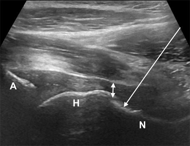
A real ultrasound of an in plane approach to a deep target, here a hip joint injection. The long bright line is a marker drawn on the image to show the needle’s path; A, H and N label the acetabulum, the femoral head and the neck. The same lengthwise approach carries a needle to a deep vessel. Image: Phey Ming Yeap and Philip Robinson, CC BY 4.0.
The first cure goes straight at the angle.
Everything here turns on one rule: the beam must meet the needle at a right angle. A beam that strikes the steel square sends nearly all its sound straight back to the probe. The needle blazes into a clean bright line. Let the beam fall slant and the sound skids off sideways, the line dimming toward nothing. The operator’s task is to build that right angle by whatever the case allows. The plainest way is to lay the needle flatter. Close to the skin, the steel runs across the beam, square to it. A shallow target allows that flat path. A deep one forces the needle to dive. The angle pays for the depth. When the needle must run steep, the probe itself is tilted to chase the angle. The near edge presses down. The far edge lifts. This heel-and-toe rock swings the beam over until it falls square on the steel, the needle brightening when the angle closes and dimming when it opens. The hand learns to rock the probe to the brightest line, reading the picture as it answers. Where the hand cannot win the angle, the machine reaches it another way. Beam steering is a quiet marvel. The probe holds a row of tiny crystals. A timed sequence of firings points the beam where the operator wants it. Tilt that aim a set number of degrees and the whole beam leans sideways, square to a steep needle. The probe stays flat on the skin. The ghost becomes a clean line at the touch of a button, no rock of the hand needed. Some machines blend several beams aimed at slightly different angles into one picture. This compound view catches a needle a single straight beam would miss. The needle faint on one angle holds bright across a fan of them. A reader on a deep line keeps the compound setting on, the extra angles working for the tip without a thought. The same reader zooms in on the trouble, narrowing the picture to the strip of tissue around the tip. The machine spends all its detail on that one band, the needle drawn larger than a wide view allows. None of this is a trick of the tip. Each is a way of aiming the sound at the steel. The steel then throws it back. Win the angle and half the battle of seeing a needle is already over, the tip riding bright on a line the eye can follow from the skin to the vessel. The whole art of brightness is the art of this one angle. Read a needle dim and the first question is always the angle, asked before any other trick is reached for. A needle that will not brighten on any rock of the probe is often a needle out of the plane entirely, the angle right and the steel beside the beam all the same. Beam steering has a ceiling of its own. The crystals can only lean the beam so far, twenty or thirty degrees, before the picture begins to suffer. A needle driven past that limit slips out of even the steered beam’s square, the brightness gone once more. The steepest sticks still ask the hand to flatten the needle as far as the target will allow.
The long axis gives the angle its best chance. With the probe run down the length of the needle, the whole shaft lies in the beam. The tip shows at the leading end of a bright line. A clean tip in long axis often shows as two short echoes lying side by side, a double mark the eye learns to read as the true end of the steel. The single surest picture of a tip is this one, the needle seen whole, its end unmistakable. A whole needle held in the long axis is the surest sight of a tip. The hold takes more skill than dropping a dot on a cross-section. The reward is a tip that never goes missing. The double mark rewards a careful eye. A needle cut at a bevel ends in a slanted face. That face throws two close echoes the screen draws as a tiny pair, the gap between them the bevel itself. A reader who knows the double tip puts the true end at the deeper of the two, never short of it. A guess that lands on the nearer echo stops a needle a bevel’s length shy of where the steel ends.
Depth works against the needle as it works against everything in ultrasound. A high-frequency probe holds a needle crisp in the first centimetres. Past its reach, a deep target loses that crispness. A reader picks the probe for the depth of the vein, then leans on the angle and the colour to claw back what depth takes away. The deepest sticks are the ones where every trick is needed at once.
A flatter approach is sometimes built into the kit. A guide clipped to the probe holds the needle at a fixed shallow angle, keeping it close to square with the beam from the start. The guide trades away some freedom of aim. In return the needle begins bright and holds that way, a fair bargain on a deep stick where the angle would otherwise fight the operator the whole way down. A guide is not free of cost. It fixes the needle’s path. A target off the planned line means pulling out and starting over. Many operators learn the freehand stick first and keep the guide for the hardest deep angles, where a fixed shallow path earns the freedom it takes.
Making the needle speak
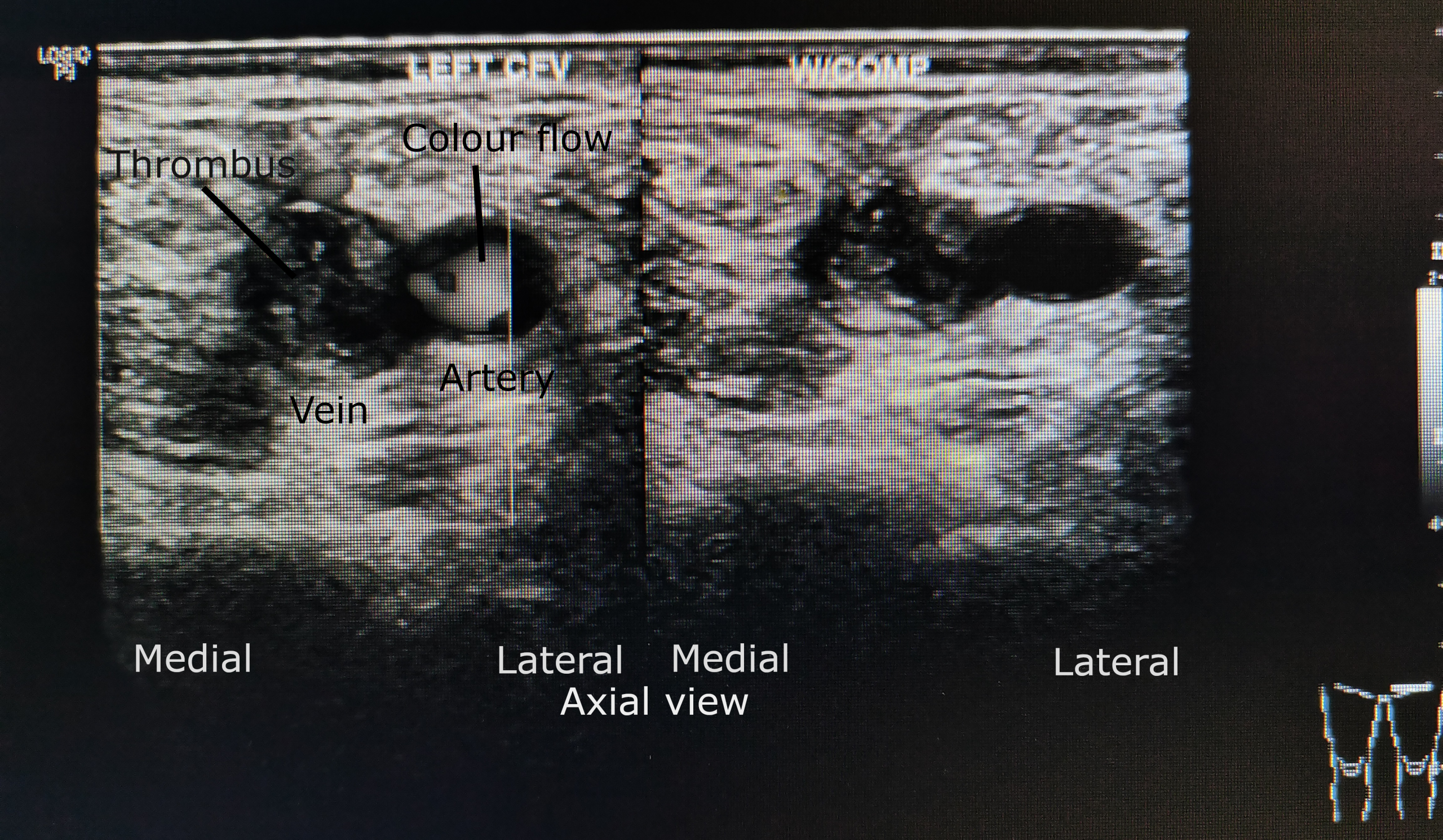
A femoral vein and artery in cross section, the colour marking the blood in the artery. The word labels were placed on the scan by its author, which was taken to show a clot in the vein. Colour like this tells an operator where an artery lies, the needle kept to the safe side of it. Image: Cerevisae, CC BY-SA 4.0.
When the angle alone will not lift the tip, the operator makes the needle announce itself. The plainest trick is often enough on its own, the angle squared and the tip back before any colour or fluid is reached for. A small jiggle of the needle sets the tissue right at its end shivering. The eye catches that flicker of motion where a still picture showed nothing, the moving tip betraying its place. This costs nothing and needs no special kit, the commonest first move when a tip goes faint. The eye is far better at catching motion than a still shape, the brain wired to notice the one thing stirring in a still field. Every trick that works by movement, the jiggle, the colour, the fluid pulse, leans on that wiring. The jiggle is a small art of its own. Too gentle and the tissue barely stirs. Too hard and the needle tears its track wider than it needs. The move is a fine waggle of the hub, a few millimetres of play that sets the tip end trembling in place. A short burst, a look, another burst, until the eye fixes the trembling point.
Colour Doppler turns that jiggle into a flag. The machine paints moving things in colour. A needle vibrated gently lights up as a patch of colour exactly at its tip. A tip lost to the grey of the tissue jumps out the instant the colour goes on. Power Doppler reads the trembling even better than colour. The colour box is kept small and laid right over the tip, just wide enough for the jiggle to show. It catches any motion at all, blind to direction. The faintest waggle of a deep tip lights it. A reader hunting a lost tip switches to power, jiggles the needle, and watches for the one spot of colour that marks the steel. The same colour guards the stick a second way. It marks the blood in a vessel. The needle is kept on the safe side of the artery.
Fluid gives the tip a voice of its own. A push of a little saline through the needle opens a small dark pocket in the tissue at its end, a bloom of black that marks where the tip sits even when the steel is hard to see. This trick, called hydrolocation, doubles as a test. A pocket that blooms inside the vein says the tip is home. A pocket that swells in the tissue says it is not yet there. The fluid earns its place twice over, the same saline that finds the tip flushing the cannula clear once the line is in. A stronger version uses agitated saline, a syringe of salt water shaken to a froth of tiny bubbles. Pushed through the needle, the froth lights up far brighter than plain fluid, a flare of sparkle that marks the tip on the dimmest screen. The same froth, sent up a central line, proves the catheter sits in the vein on its way to the heart. Hydrolocation asks for a clear fluid and a measured push. A drop too small shows nothing. A flood pushes the tissue apart and hides the tip in its own pocket. A breath of saline, half a millilitre, blooms just enough to mark the end and no more. The bloom fades in a moment. The reader watches for it at the instant of the push.
The needle itself can be built to be seen. An echogenic needle carries microscopic pits or facets cut into its last centimetres. The choice of needle is made before the stick, not in the middle of it. These catch the beam and scatter it back even from a steep angle. A smooth needle loses that brightness on a steep dive. The textures come in a few designs. Some needles wear rings of tiny grooves near the tip. Some carry a corner pattern that throws the beam straight back from any angle. Some hold a coat that scatters the sound. Each buys back the brightness a plain needle gives up, the gain largest exactly where a plain needle fails. The newest machines hunt the tip on their own. A processor trained on thousands of needles marks the tip with a coloured ring that follows the needle in, a steady label where the eye once strained. No single trick wins every tip. A deep steep needle in a busy field can defeat the angle, the jiggle, and the colour one at a time. The same needle shows clean when two are used at once, the beam steered and the needle jiggled together. A reader stacks the tricks on the hardest tip until the steel cannot hide. An echogenic needle costs more than a plain one. The extra pays for itself on a deep target set at an awkward angle. There the tip stays readable at a depth that swallows a plain needle.
What no setting can replace
Under all the tools sits one discipline no button can supply: never advance a tip you cannot see. The instant the line fades, the rule is to stop the needle, recover the tip by whatever means, and only then push on. A tip lost for a moment is a warning to heed. The hand stops at once. The operator who obeys that one rule turns every trick above into a safety net. The same tricks go to waste for an operator who forgets it. The rule sounds plain. Pressure is where it breaks. A code is running, the vein is deep, the hand wants to push for the flash of blood. In that moment a tip slips away unseen, a back wall an easy mistake. The operators who never harm a patient on a deep stick are the ones who stop every time the tip fades, with the room watching and the clock running. The tricks also come in an order a reader keeps by heart. Square the angle first. Jiggle and watch next. Reach for the colour if the jiggle is not enough. Lay down a breath of saline if the colour still leaves a doubt. The order keeps the slowest trick in reserve for the tip that will not show any other way.
On a handheld
The handheld carries the same toolkit into a pocket. The probe rocks heel to toe for the angle just as a cart’s does. The colour Doppler that flags a jiggled tip runs on the phone. The light body of a wireless probe makes the fine heel-and-toe easier than a heavy cabled head, the smallest tilt of the wrist answered at once. The fine control matters more on this task than on any other scan. Reading a tip means rocking the probe by a single degree to find the bright line, then holding it dead still as the needle advances. A heavy probe fights the hand. A cable dragging on it does the same. Either loses that one degree of tilt the bright line lives in. A line goes in at a bedside with the tip held bright the whole way down, no procedure room and no cart required. The phone screen reads a faint tip even in a lit room, the colour and the gain set with a fingertip. A reader who loses a tip taps up the gain or flicks on the colour, the controls under the same thumb that steadies the probe.
Some of the toolkit asks more of a small machine than others. Beam steering and the cleanest colour need the processing a good handheld now carries. A clinic choosing a probe for guided access does well to check that it steers its beam and runs a clean colour, the two settings that rescue a fading tip when the hand alone cannot. The saved image on the phone records the tip in the vessel, proof for the chart that the line went where the eye said it did. A saved clip of a tip blooming inside a vein is more than a record. It shows the next learner what a true tip looks like: the dark bloom of saline, the spot of colour, the double mark of the bevel, caught in one short loop to study away from the bedside. The methods do not need the cart. They need a probe that steers a beam, a screen that shows a clean colour, and a hand that knows the order to try them in. A trainee on a ward rehearses the angle, the jiggle, the colour, and the fluid on real veins, the lesson shared on one phone screen. A clinic far from a procedure suite holds the whole of guided access in a handheld, the same skill a teaching hospital teaches placed in the hands of a nurse on a remote ward. A line whose tip was watched into the vein and saved as proof rarely needs the chest film a blind stick once called for. The cost of the tool has fallen as far as its size, a guided line no longer the preserve of the hospital that can afford a cart.
Common questions about needle tip visualization
Why is the needle tip hard to see on ultrasound?
Because ultrasound reflects best off a surface it strikes square. A needle diving toward a deep target meets the beam at a slant that throws much of the sound away from the probe. The tip is also the smallest part of the needle. It vanishes the moment the needle tilts out of the thin plane of the beam.
What is hydrolocation?
It is the use of a little fluid to find the tip. A small push of saline through the needle opens a dark pocket in the tissue at the tip, marking its position even when the steel is faint. A pocket that blooms inside the vein confirms the tip is in the lumen.
How does colour Doppler help see the needle?
It paints movement in colour. A gentle jiggle of the needle makes the tissue at its tip vibrate. The colour lights up that spot, pulling a lost tip out of the grey. The same colour shows the blood in nearby vessels. The needle is kept on the safe side of the artery.
What is an echogenic needle?
A needle textured with tiny pits or facets near its tip. The roughened surface scatters the beam back to the probe even at a steep angle. A smooth needle loses that brightness on a steep dive toward a deep target. An echogenic needle costs more than a plain one. Hard sticks are where it pays for itself.
Can a handheld ultrasound show the needle tip clearly?
Yes, with the same methods a cart uses. The probe is rocked to square the beam on the needle, the tip is jiggled under colour Doppler, and a little saline marks its place. A light wireless probe makes the fine probe tilt easier. A good one steers its beam and runs a clean colour for a fading tip.
About the Author
Julien Mercier
Senior R&D Engineer · Medical Ultrasound Transducer Development
Senior R&D Engineer with an M.S. in Applied Physics and over 15 years of experience in medical ultrasound transducer development, specializing in the design verification and performance testing of high-frequency imaging transducers. Currently leading the development and verification of the company’s next-generation high-frequency linear-array transducer, responsible for imaging performance evaluation and reliability analysis in preclinical testing. Brings extensive hands-on experience in piezoelectric element tuning, beamforming parameter optimization, and system-level performance testing.