Locating a filler embolism with ultrasound is how a clinic turns a vascular emergency from guesswork into a targeted fix. The probe shows the trapped filler as a dark pocket and the stopped flow in the vessel beyond it. It guides the antidote into the deposit, then watches the blood return.
The emergency behind the scan
A hyaluronic acid filler can enter an artery during an injection. The filler blocks the vessel. The skin that artery fed starts to lose its blood supply. Near the eye the same block can reach the vessels of the retina. This is a vascular occlusion, the emergency every filler injector prepares for.
An occlusion does not need a careless injector. It can follow a correct technique in a risky spot. A small artery runs where the needle goes. A bolus delivered under pressure enters it. The danger sits highest at the glabella, the nose and the nasolabial fold, where the arteries are small and connect toward the eye. The bolus that does the harm is often small. It takes only a little filler forced into the lumen of a facial artery under thumb pressure to seed a block that travels. Slow injection at low pressure lowers the odds. A blunt cannula lowers them further.
The antidote is an enzyme, hyaluronidase, that breaks down hyaluronic acid. Placed into and around the trapped filler, it dissolves the plug and lets the blood move again. The enzyme acts only on hyaluronic acid, so it suits this kind of filler. It works best when it reaches the deposit fast and sits right inside it. A first amount starts the job. The clinician usually gives more over the following minutes, since a large plug takes repeated doses to break apart. The whole aim of the emergency is to get the enzyme to the deposit before the tissue downstream dies. Every minute the plug stays in place is tissue the patient may lose. The enzyme is the only thing that reverses a hyaluronic acid block. Pressure, warmth and tapping have all been tried. None clears a plugged artery the way the enzyme does. The whole emergency turns on getting it to the right spot before the window closes.
Speed decides everything. The skin has hours. The eye has minutes.
Reading the first signs
An occlusion announces itself. The skin over the blocked vessel turns pale as the blood leaves it. A dusky mottling follows. The patient feels pain out of proportion to the procedure. Pressing the skin and watching the color crawl back shows the supply is cut. The pain is the clue that gets missed. A patient expects some soreness from a needle, so an ache out of proportion to the work can pass for ordinary until the skin colors. A clinician who treats unexpected pain as a warning, not a nuisance, gains the early minutes that matter.
The signs run in a sequence. Reading the stage tells the clinician how much time is left. The pale skin and the pain come first, within minutes to an hour of the injection. A net-like pattern spreads over the next hours as the starved tissue struggles. Small blisters can rise on the surface after that. A dark crust forms where the skin has died, days later, if the supply never came back. Catching the problem at the pale stage gives the best chance of saving the tissue. Each later stage means more of it is already gone. The clock starts at the injection, not at the first dark patch. A clinician who treats the moment the skin pales buys back hours that the one who waits to be certain loses for good.
The signs alone do not say where the filler sits or how much is there. A bruise can mimic an early occlusion. Swelling can hide the pattern. The clinician who suspects one needs to know fast whether filler is sitting in or against a vessel, and where it sits. This is the question the scan answers.
What the scan shows
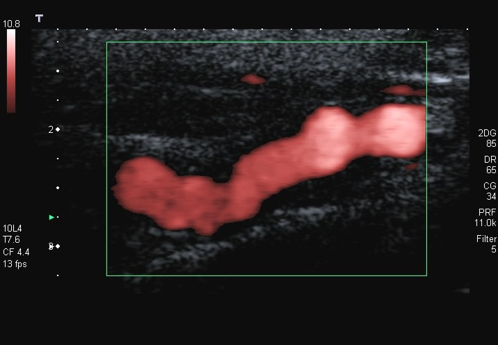
Power Doppler fills a flowing artery with color, the signal a clinician watches in a filler occlusion. A blocked vessel shows no color beyond the plug. A return of color says the enzyme cleared it. This is a power Doppler example on a neck artery. It is not a filler case. The box and the settings are the machine’s own.
Hyaluronic acid filler has a clear look on ultrasound. It reads as a dark pocket that looks like fluid, often with a brighter edge around it. Old filler placed months earlier can sit deeper than the surface suggests, or further from the spot the needle entered. The probe finds these pockets and reads their size and their depth. It tells one pocket from several. It shows whether a deposit sits in the path of a vessel or off to the side. Each of these readings shapes where the needle goes next.
Color Doppler adds the flow. A healthy artery fills with color as the blood pulses through it. An artery blocked by filler shows no color past the block. The clinician runs the probe along the vessel to find the point where the flow stops.
Read together, the gray image and the color flow place the occlusion with a precision the surface never offers. The clinician sweeps the gray scale first and marks every dark pocket of filler, noting its size, its depth, and how far it sits from the entry point, since a bolus pushed under pressure can track along a plane and lodge a centimeter from where the skin went pale. Color Doppler then follows the artery that feeds the pale skin. The point where the color stops marks the head of the block. The two readings line up: the filler pocket sits at or just upstream of the dead end in the flow. The same sweep separates the things that imitate an occlusion, a pocket of pus, a spreading bruise, a vein swollen with blood, each of which reads its own way and calls for its own response. By the end of a careful scan the clinician knows what is blocking the vessel, where it sits, how deep the needle must travel to reach it, and which way the flow runs around it. That picture is the plan.
Telling these apart changes what happens next. A lump of filler reads differently from pus or a bruise. The treatment for each is different. The scan turns a guess about what is happening under the skin into a picture of it. Pus shows as a pocket with debris that shifts under the probe. A bruise spreads without a sharp edge. A swollen vein fills with color on Doppler where the filler stays dark. Reading the deposit correctly points the clinician to the enzyme, not to an antibiotic or a wait that would cost the skin.
The deposit does not always sit where the symptom shows. The skin can go pale in one spot while the plug sits a short way off. A scan of the whole area finds where the product settled.
Guiding the antidote to the deposit
Blind treatment floods a whole area with enzyme and hopes some of it reaches the filler. Much of it does not. Ultrasound changes the aim. The clinician sees the filler pocket on the screen. The needle lines up with the beam so its shaft shows as a bright line. The tip goes into the deposit under live view. For a larger plug the clinician moves the tip through it in passes, spreading the enzyme across the whole deposit. The screen shows the pocket soften and shrink as the enzyme works. Reported cases place the enzyme this way with a fine insulin needle and watch the tissue around the deposit fill with flow within minutes. The needle stays in the plane of the beam so its full length shows, since a tip lost outside the plane places the enzyme blind even with the deposit in view. The clinician moves the tip through a larger plug in passes, laying the enzyme across the whole deposit, not into one corner of it.
The lower dose carries its own value. Hyaluronidase dissolves the filler that caused the problem. It also dissolves the filler placed for the treatment itself. A targeted injection clears the occlusion while sparing more of the rest. Seeing the deposit makes that precision possible. A blind flood of enzyme dissolves the good filler placed weeks before, undoing the treatment the patient paid for. The guided injection takes out the plug and leaves the rest, so the rescue does not turn into a second loss.
How much enzyme, and how often
The amount of enzyme is a judgment, not a fixed recipe. Blind protocols lean on large doses, repeated every hour, because the clinician cannot see whether the enzyme is reaching the filler. A guided injection changes that. One reported case placed around 150 units straight into the deposit with a fine needle and saw the tissue around it fill with flow within minutes. Seeing the target lets the clinician start with what the deposit in front of them seems to need, then judge the next dose from the screen.
The dose repeats on the evidence of the scan. The clinician places the enzyme, watches for color to return, and gives more if the vessel stays empty. A large plug can take several rounds to break down, since the enzyme works on the surface of the filler it touches. The deposit softens and shrinks across the rounds, which the gray image shows directly. The endpoint is flow restored along the vessel, not a number of units reached. A clinic without the scan repeats on the clock and the look of the skin, dosing every hour and hoping. The guided clinic doses on the vessel itself, stopping when the flow returns and not before.
The enzyme acts only on hyaluronic acid. A filler made of another material, a calcium-based or a permanent one, does not dissolve this way. The scan that shows the deposit as a dark pocket of fluid supports the choice to reach for the enzyme. For those other fillers the emergency runs a different course, one the enzyme cannot rescue.
Watching the blood come back
The scan does not stop when the enzyme goes in. Color Doppler shows whether the flow returns. The clinician watches the vessel beyond the block for color to fill it again. A return of flow says the enzyme reached the plug and the supply is coming back. An empty vessel says the work is not done. This signal is the part blind treatment never has. A clinician working without the probe judges by the skin color and the clock, both of which lag behind what the vessel is doing. The Doppler reads the vessel itself, in the moment, so the next decision rests on what the flow shows.
The return can take more than one round. A first dose can open the vessel partway. The clinician gives more enzyme to the same deposit and checks again. The flow builds back over the rounds. Each round is a question the screen answers: is the vessel open yet. The clinician stops when the color runs the length of the vessel, not when the syringe is empty.
The check guards against stopping too soon. The skin can look better before the vessel is fully open. A scan that shows flow restored along the vessel gives a firmer endpoint than the surface color. It also documents that the supply came back.
After the flow returns
Restored flow is the turning point, not the finish. The skin downstream that was starved needs watching over the hours and days that follow. A patch that stayed pale too long can still break down, so the clinic follows the area and sometimes scans it again to confirm the vessel stays open. A deposit cleared only in part can block the vessel a second time as the swelling around it shifts. The first day is the one to watch closest. A vessel that reopened can close again. Skin that looked saved at the clinic can darken overnight. The clinic that sends the patient home with a number to call and a low bar for returning catches the second event in time.
The scan guides those days. A repeat look shows whether the dissolved filler has cleared or whether a remnant still presses on the vessel. The clinician can place more enzyme on a remnant the screen still shows. The patient leaves with a plan built on what the vessel is doing, not on hope that the worst has passed.
Scanning without losing time
The scan must not slow the treatment. A clinician who suspects an occlusion gives hyaluronidase without waiting for a perfect image, because the clock is the enemy. The scan runs alongside the treatment. The first dose can go in on the clinical signs. The probe then refines where the next dose lands. The order is fixed: the drug goes in on suspicion, the scan runs beside it. A clinician who stops to perfect the image before injecting has misread the clock. The probe speeds the rescue. It must not delay it.
A clinic ready for this has the pieces in place before the day it is needed. The hyaluronidase sits within reach, in date, in a known drawer. The probe is charged and paired to its screen. The staff know the signs and the steps without looking them up. The scan adds its value only inside a plan that was rehearsed. A probe bought and left in a cupboard helps no one at the moment it is needed. The drill is what turns the equipment into a response.
The map from before the needle
A clinic that scanned before the injection starts the emergency ahead. The map made going in already shows where the arteries run in this face and how deep they sit, so the search for the deposit is shorter when minutes matter. The clinician knows which vessel is likely blocked before the probe even touches the skin. The same probe and the same screen serve both jobs. The device that mapped the vessels going in is the one that finds the deposit and guides the enzyme coming out. A clinic set up to map its injections is, by the same equipment and the same habit, set up to manage their worst complication. The records help here too. A clinic that saved the map from the injection can pull it up mid-emergency, matching the pale skin to the artery that feeds it. The minutes saved by knowing the anatomy already are the minutes the skin has left.
What ultrasound does not replace
Ultrasound does not replace the emergency protocol. The clinic still needs the enzyme on hand, a rehearsed plan, and the judgment to act fast. The scan guides the treatment. A clinician who cannot read the image gains nothing from owning the probe. The probe earns its place only in hands that have rehearsed the whole drill: the drug ready, the scan practiced, the repeat understood. The tool sharpens a prepared response. It cannot supply one.
An eye emergency sits beyond what a facial scan handles. Filler that reaches the artery behind the eye threatens sight within minutes and calls for immediate specialist care, often aimed at the eye itself. The probe has its role in the face, finding and clearing a deposit there. It does not open a blocked retinal vessel. Knowing where the probe stops is as much a part of using it well as knowing what it can do. The injection that might save the eye goes behind the eye, on a clock of minutes, in a specialist’s hands. The facial probe plays no part in that step. A clinic that reads the face well still has to recognize the moment the danger has moved past the face.
Common questions about filler occlusion ultrasound
How does ultrasound help in a filler vascular occlusion?
It shows the trapped filler as a dark pocket and shows the flow stop on color Doppler. The clinician then injects hyaluronidase straight into the deposit under live guidance and watches the blood return. Seeing the filler means a smaller dose of enzyme aimed at the deposit in place of a blind flood.
What does the filler look like on the scan?
A hyaluronic acid filler reads as a dark pocket that looks like fluid, often with a brighter edge. Color Doppler shows a blocked artery as an absence of flow beyond the deposit. The clinician finds the point where the color stops and matches it to the pocket on the gray image.
Does guiding the enzyme by ultrasound use less of it?
Yes. A blind injection floods a whole area and hopes the enzyme reaches the filler. A guided injection places it straight into the deposit. Reported cases clear the occlusion with far less enzyme this way, which also spares more of the filler placed for the treatment itself.
Should the scan come before giving hyaluronidase?
No. The drug should not wait for a perfect image. A clinician who suspects an occlusion gives hyaluronidase on the clinical signs and runs the scan alongside the treatment. The probe then refines where each further dose lands. The scan supports the treatment. It does not hold it up.
How does the scan confirm the treatment worked?
Color Doppler shows the flow returning in the vessel beyond the block. A return of color says the enzyme reached the plug and cleared it. An empty vessel says more is needed. The check guides whether to repeat the injection and gives a firmer endpoint than the skin color alone.
Is speed that important?
Yes. The skin downstream of a blocked artery has hours before the damage is permanent. An occlusion near the eye is measured in minutes. Anything that shortens the time from the first sign to the enzyme in the right place improves the outcome, which is what the scan does.
Does ultrasound handle an eye complication?
No. Filler that reaches the artery behind the eye threatens sight within minutes and needs immediate specialist care. The probe has its role in the face, finding and clearing a deposit there. It does not manage the eye itself. Knowing that limit is part of using it well.
About the Author
Julien Mercier
Senior R&D Engineer · Medical Ultrasound Transducer Development
Senior R&D Engineer with an M.S. in Applied Physics and over 15 years of experience in medical ultrasound transducer development, specializing in the design verification and performance testing of high-frequency imaging transducers. Currently leading the development and verification of the company’s next-generation high-frequency linear-array transducer, responsible for imaging performance evaluation and reliability analysis in preclinical testing. Brings extensive hands-on experience in piezoelectric element tuning, beamforming parameter optimization, and system-level performance testing.