


































Canine Feline Renal Failure Ultrasound Handheld Assessment
A renal ultrasound reads the kidneys of a dog or cat already in failure, looking past the failing numbers to the kidney itself, its shape and its substance. The bloodwork names the failure first, reading the waste the kidneys can no longer clear. What the scan adds is the other half of the story, the state of the organ behind the failure, which points to how far back the damage goes and how much of it might still turn around.
What the scan cannot do
The scan cannot tell how well a kidney is working. Function is a chemistry, the rate at which a kidney clears waste from the blood. No picture can measure a rate. The blood creatinine, the SDMA, the concentration of the urine: these read function, and read it long before the kidney looks much wrong on the screen, where a kidney with half its work gone can still pass for normal. Structure and function part ways here, the shape lagging behind the chemistry that has already turned.
This catches owners out. The screen often shows a kidney close to ordinary even in deep failure, the damage held in the blood, the shape barely touched. Owners find that hard to credit of an organ said to have failed. Early failure under a near-normal scan is an everyday sight.
The scan’s job lies past the diagnosis. It takes up the questions the bloodwork cannot answer. Is one kidney to blame, or both. Has the kidney shrunk and scarred over years? Or swollen in a matter of days? Is something blocking it that a surgeon could clear. These are the scan’s questions. Their answers steer all that follows.
Why the animal came in
The animal rarely arrives named as a kidney case. It comes in drinking and passing far more water than it should, a thirst the owner has watched grow for weeks. It comes off its food, dropping weight, the coat gone harsh and staring. An older cat losing weight and emptying every water bowl in the house is the picture every vet learns to scan for. A cat comes in quietly fading, the commonest patient of all for failing kidneys.
By the time these signs show, the kidneys have spent much of their reserve. Half the working tissue can be gone before the blood creatinine climbs at all, so the disease is often well advanced by the day it is finally named. The thirst and the wasting come of a kidney no longer holding water back or clearing waste, the body flushing through what the kidney leaks. The blood test that follows reads the failure, the scan booked to read the kidney behind it.
Some arrive sicker and faster. An animal that has stopped eating, gone dull, begun vomiting over a day or two, may be in an acute crisis. The scan is run urgently then, hunting for the blockage or the poison behind a fast failure.
The healthy kidney as a baseline
Every reading starts from the look of a normal kidney. A healthy one holds a clean bean shape, a smooth outline, and a size that fits the animal. Inside, two layers show plainly: an outer cortex of an even mid-grey, and within it the darker pyramids of the medulla, where the urine gathers. The line between the two, the corticomedullary junction, stands out as a sharp line in health. The centre, where the urine collects before its run to the bladder, reads as a bright line holding no pocket of fluid. The whole organ in a cat runs to around the length of a thumb, give or take, a size the eye learns by long looking.
A cat’s kidneys run a little smaller than a dog’s. They read a shade brighter as well, and a little fat around them can lift the cortex brighter still. Knowing the normal range for the animal on the table is what keeps a healthy kidney from being called a sick one.
How the kidney is scanned
The kidneys are not the easiest organs to reach. The left sits high under the last ribs on its flank, the right tucked further forward still, half-hidden behind the liver and the rib edge. The animal is laid on its back or its side, the flank clipped and gelled, the probe angled up under the ribs to bring each kidney whole onto the screen.
Each kidney is read in two planes. The long axis shows the bean shape end to end, the cortex and medulla in their layers. The probe is turned a quarter circle for the short axis, a round cross-section that confirms what the long view showed and catches anything hiding in it. A kidney read in one plane only is half read.
Reaching the kidneys can take patience. They lie deep in a big or barrel-chested dog, shadowed by gas and the ribs. Such a patient may need rolling, the probe worked in between the ribs to bring a kidney whole onto the screen. The work goes quickest on a small animal whose kidneys sit near the surface.
Measuring the kidney
Numbers pin down what the eye estimates. The kidney is measured along its length, the figure set against the range known for a cat or for a dog of that build. A cat’s kidney keeps a fairly tight normal length, so a measurement under it carries real weight, the shrinkage of chronic disease put to a number on the page. The two kidneys are measured against each other as well, one much smaller than its partner having lost ground the partner has held.
The brightness is judged by comparison. The cortex is read against the liver lying beside it, since a cortex brighter than that liver is a cortex turning to scar. The width of the collecting space is measured as well, read for anything from a thread of fluid, a mild backing-up, to a space ballooned wide by a blockage below. Even a sliver of fluid in the pelvis is noted down, since it can be the first whisper of a block forming downstream.
The shrunken, scarred kidney
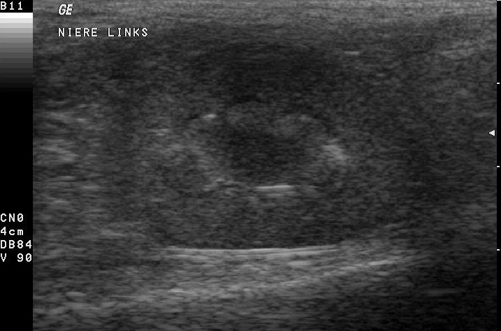
Chronic kidney disease leaves a kidney small. Years of slow scarring shrink the organ, pull its outline into bumps and hollows, and rob it of the clean shape it began with. A kidney plainly undersized for its animal is one with years of disease behind it.
The picture of a chronic kidney runs through the whole organ. The cortex turns bright, brighter than the liver or the spleen it is read against, the grey of healthy tissue given over to the white of scar. The clear line between cortex and medulla blurs and fades, the two layers bleeding together where they once stood apart. The outline loses its smooth curve, notched where wedges of tissue have died and drawn in. Small pockets of fluid open in the centre, a mild swelling of the collecting space, the kidney no longer holding its old form. In a cat, both kidneys often carry this look at once, shrunken, bright, and lumpy, the quiet end of the disease that fells more old cats than any other illness. Across a kidney small for its animal, these signs together spell a long, settled disease that no treatment will turn back.
The scarred kidney is the hard news the scan delivers gently. It tells an owner the failure is the slow, lifelong kind, a kidney worn out across years. There is no operation for it, only the long care that eases its course and buys good time.
Both kidneys usually share the look. Chronic disease works on the pair together, since whatever drives it, the slow loss of age, a long inflammation, a breed’s inherited flaw, reaches both through the same blood. Sometimes only one kidney is hit. A local injury, an old dead wedge or a scar, shrinks and dims that single organ. Its partner stays normal and carries the work of both. An infarct, a piece of kidney killed when its small artery clotted, leaves just such a mark, a sharp notch in the outline over a pale triangle of dead tissue.
A scarred kidney is read against the calendar. A scan today, set beside one from a year back, shows whether the kidney is holding steady or wasting on, the pace of the disease laid bare in the change between two pictures.
The swollen kidney
A sudden injury swells a kidney. An acute insult, a poison, a collapse of the blood supply, an infection raging through it, leaves the kidney big, tense, and swollen. A big kidney on a newly sick animal speaks of something sudden. The cortex may brighten as well, the swollen tissue turned white, the kidney straining against an injury only hours or days old.
Antifreeze is the classic poison here. A dog or cat that has licked spilled antifreeze grows a pair of big, painful, oddly bright kidneys inside a day, the ethylene glycol laying down crystals through the tissue. The scan can show a striking brightness, a halo of white through the kidney, that points hard at the poison when the history is a blank. A rim of fluid gathering around the kidney can show in the worst of these cases, the organ weeping under the assault.
The swollen kidney often carries hope. Caught in time, an acute injury can be fought, the kidney rested and supported as it heals, dialysis buying days in the worst of it. A kidney swollen today may read close to normal in a month, the failure behind it hauled back from the edge. The line between a kidney that will come back and one that will not is often drawn only in the weeks after, the scan repeated to watch the swelling settle or the damage set.
A swollen kidney is a kidney under pressure from a block below it. The scan follows the swelling down the ureter toward the bladder, hunting the stone or the narrowing that dammed the flow. A kidney swollen on one side sends the search down that one ureter.
What the scan answers
The blood says the kidney is failing. Why it is failing is the scan’s to answer.
The blocked kidney
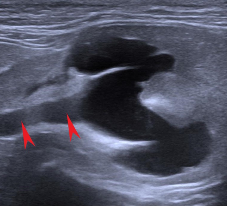
Of all the things the scan looks for, a blockage is the one it hopes above all to find. A blocked kidney is a kidney that can often be saved, the one cause of failure a surgeon can often undo.
A stone or a narrowing in the ureter, the tube from kidney to bladder, dams the urine behind it. The pressure backs up into the kidney and swells the collecting space into a balloon of fluid, the picture called hydronephrosis. The scan reads the ballooned centre, follows the ureter down toward the stone or the kink doing it, and measures how far the harm has run. The wider the balloon and the thinner the rim of kidney left around it, the longer the block has sat and the less there is to save. A kidney can swell to many times its size when a block sits long enough, a thin shell of tissue stretched around a bag of trapped urine.
Cats are the ones this strikes hardest. A cat’s ureter is finer than a thread, fine enough for a stone no bigger than a poppy seed to block it, the kidney swelling in silence behind. A cat in sudden failure has its ureters hunted along their whole length, since a blocked one found early is a kidney still there to save. Such a stone may have sat in the kidney for years. The day it slips into the ureter and jams is the day the trouble starts.
Time is the whole of it. A blocked kidney can recover if the block comes off within a day or two. Each week it stays dams more of the kidney dead. The scan that finds a block is read with the clock already running.
Reading the two kidneys together
A kidney is never read on its own. The two are held against each other, since the pair says more than either does alone. Two kidneys failing together, both small, both bright, speak of a disease running through the whole animal. A single ruined kidney next to a healthy one speaks of a lone organ lost, the load fallen to the kidney that remains. Two kidneys that look well on an animal whose blood has failed pose the hardest question, the damage still beneath what the picture can show.
The lone working kidney is a finding to guard. An animal living on one kidney has no margin to spare, so that kidney is watched close and spared every avoidable harm, the wrong drug, the missed blockage, the untreated infection. A young animal born with a single kidney, or one left after the other came out, lives well enough on it, the lone organ grown large to manage the load.
What else the scan turns up
A kidney scanned for failure often gives up more than the failure. Cysts are common, round pockets of clear fluid in the cortex. In a breed prone to the inherited kind they crowd the kidney in their hundreds and become the disease itself, squeezing the organ out over years. A single large cyst can grow big enough to press on the working tissue around it, the cyst itself turning into the problem.
Masses turn up too. A growth in a kidney raises the question of a tumour, a solid lumpy mass breaking the even grain of the tissue. The scan measures it and reads the other kidney and the belly for any spread before a needle is ever put in. A lymphoma can swell both of a cat’s kidneys at once, a bloated look that passes for plain failure until the needle names it.
Stones sit in many a failing kidney. A bright stone with its dark shadow, lodged in the collecting space, may be the cause of the trouble or an old bystander. The scan weighs which by the kidney behind it. A swollen kidney behind the stone marks it as the active block. Without that swelling, the stone is likely an old passenger, formed long ago and quiet since.
The scan rarely reads one thing alone. A kidney scanned for failure shows its stones, its cysts, the state of the bladder downstream, all in the same sweep. Each finding is weighed for whether it drives the failure or merely rides along with it.
Doppler and the blood flow
Beyond the kidney’s shape on the grey picture, colour Doppler reads its blood. Laid over the kidney, the colour paints the vessels branching through the tissue. From those vessels comes a number, the resistance the blood meets on its way through. That resistance climbs in a kidney that scarring has stiffened, its vessel tree thinned, the flow labouring through hardened ground. The number is read the same way each time, the same vessels sampled in the same spot, so one scan can be set against the next.
The number is no diagnosis on its own. Resistance reads high in a kidney that scar has stiffened. The reading is weighed alongside the shape and the bloodwork, never trusted by itself. A single reading counts for little. A resistance climbing across two scans, months apart, tracks a kidney stiffening over time.
A single Doppler reading is a snapshot that means little alone. The number told across scans, climbing from one year to the next, traces a kidney that scar is slowly stiffening, its vessel tree thinning as the disease wears on.
Following the disease over time
Kidney disease is a long road. The scan is one of the markers set along it. A cat newly found in early failure has its kidneys scanned as a baseline, the shape and size and brightness written down for the scans to come. The first picture is the one every later scan is measured against, so it is taken with care and saved in full.
Each later scan is read against the first. A kidney shrinking and brightening from one scan to the next is a disease pressing on, the cue to tighten the care around it.
The scan also catches the turns. A chronic kidney that suddenly swells, or a fresh pocket of fluid that opens in its centre, says something acute has landed on top of the old disease, an infection or a new blockage to chase down fast. An old kidney case that drops suddenly is scanned at once, the new trouble on top of the old the thing likeliest to be fixable.
None of this stands in for the bloodwork. The blood and urine carry the routine watch, read often, perhaps monthly. The scan comes a few times a year, called in when the numbers jump or the animal turns. Each catches what falls between the other’s readings.
A scan kept the same way each time is a scan that can be trusted across the years. The same windows, the same measurements, the same comparison to the liver: the steadiness is what lets one year’s picture be read honestly against the last.
When the picture and the blood disagree
The scan and the bloodwork do not always tell the same story, the gaps between them carrying their own meaning. A normal-looking kidney on an animal with failing blood numbers is the common one, the damage finer than the picture can catch, a reminder that a clean scan does not clear a kidney.
Sometimes the picture runs ahead of the blood. It shows a shrunken, bright, scarred organ weeks or months before the numbers give the failure away. That kidney is coping for now, its reserve thinning. The animal is watched closely, the scan having seen the trouble before the blood would own it.
The blood and the scan answer different questions, and a full reading of a kidney case wants both, the numbers and the pictures set side by side and weighed as one.
What the scan changes
A scan turns the plain fact of failure into a course of action. The same blood result, kidneys failing, sends one animal to theatre, another to the drip, a third home with a diet sheet. A blockage found sends the animal to surgery. An acute swollen kidney earns a place on the drip and a bed in the ward. Two kidneys worn small turn the talk toward the long care, the diet and the drugs that lighten a kidney’s load.
It guards against the wrong move as well. An animal in failure cannot clear drugs the way a well one does. A scan that reads the kidneys badly scarred warns the vet to lighten or drop the medicines that would build up and poison. A pocket of pus the scan picks out, an abscess walled in the kidney, sends the animal toward drainage and a hard course of antibiotics.
It sets the honest expectation, too. A worn, shrunken pair tells an owner the road ahead is a long decline, a hard kindness, the truth that lets a family plan and a vet aim the care where it can still do good.
The scan also sets the pace of the watching. A kidney on the move pulls its animal back for the next scan sooner, the schedule tightened to match a disease that has begun to press.
The rest of the story
A renal scan earns its keep in what it rules in and rules out. It tells a vet whether a failing kidney is a kidney worn down over years or one struck this week, a difference that sets the whole plan.
What the scan cannot give is the function itself, the job the bloodwork does. The scan reads the organ behind the numbers, the shape and the substance the blood cannot show, the two together telling more than either alone.
The picture and the numbers tell one story between them. A vet weighs them together over the visits, the kidney’s shape on the screen set against its failing chemistry, until the course of the disease comes clear.
In a failing kidney where the bloodwork has read the loss already, the scan is there for the rest of the story: how far the kidney has fallen, what drove it down, and whether the fall can still be stopped.
Common questions about renal ultrasound
Can an ultrasound diagnose kidney failure in a dog or cat?
No, not on its own. Kidney failure is a loss of function, read by blood and urine tests, the creatinine and the SDMA. The scan’s job is the structure behind that function: it tells why the kidneys are failing and whether the cause can be treated, alongside the bloodwork that names the failure.
What does a kidney with chronic disease look like on ultrasound?
Small, with a bright cortex and a blurred line between its inner and outer layers, often with an irregular, dented outline. In cats both kidneys usually share the look. These signs together, on a kidney small for the animal, point to long-standing disease.
Can ultrasound tell acute kidney failure from chronic?
Often, yes. Chronic disease leaves a kidney small, its cortex bright, its outline scarred and dented over long years. The acute kidney has not had that time: it reads large, tense, freshly swollen. That difference decides the outlook. An acute kidney can sometimes recover its work over the weeks that follow, a recovery scarring never allows once it is laid down.
Why is a blocked kidney important to find?
A blockage, usually a stone caught in the ureter, dams urine and swells the kidney behind it. Caught early, within a day or two, the blockage can be cleared and the kidney saved. Each week it sits, more of the kidney dies behind the dam. The scan finds the block and the swelling, and the clock starts the moment it does.
How often should a pet with kidney disease be scanned?
A baseline scan at diagnosis, then a repeat now and then to track the kidneys over time, reads how fast the disease is moving. A sudden change in the animal earns a scan sooner, to catch anything acute landing on top of the chronic disease. The bloodwork carries the routine monitoring between the scans.

About the Author
Julien Mercier
Senior R&D Engineer · Medical Ultrasound Transducer Development
Senior R&D Engineer with an M.S. in Applied Physics and over 15 years of experience in medical ultrasound transducer development, specializing in the design verification and performance testing of high-frequency imaging transducers. Currently leading the development and verification of the company’s next-generation high-frequency linear-array transducer, responsible for imaging performance evaluation and reliability analysis in preclinical testing. Brings extensive hands-on experience in piezoelectric element tuning, beamforming parameter optimization, and system-level performance testing.