Facial Blood Vessel Mapping With Ultrasound Before Hyaluronic Acid Filler
Facial blood vessel mapping before a filler injection is the search for the arteries a needle must stay clear of. A few of those arteries connect, through hidden routes, to the artery behind the eye. Those few are the ones a misplaced bolus can blind a patient through. They decide where the needle takes the greatest care.
The route to the eye
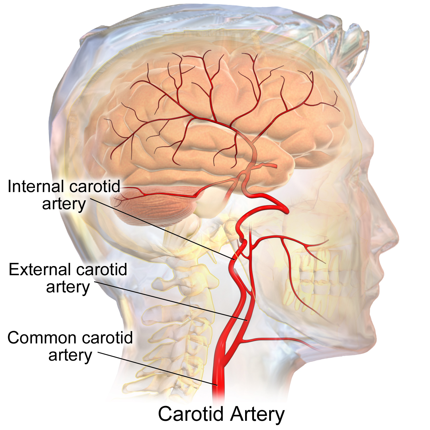
The two arterial systems behind the danger. The internal carotid feeds the eye and the brain. The external carotid feeds the face. They join near the eye, which is how filler in a facial artery can travel back toward the retina. This is a medical illustration. It is not an ultrasound image. The labels are part of the original.
The face draws its blood from two systems. The external carotid artery feeds much of the face through the facial artery and its branches. The internal carotid artery feeds the eye through the ophthalmic artery. It also sends small branches onto the forehead and the nose. The two systems meet. Around the inner corner of the eye, branches of the external carotid join branches of the internal carotid.
That meeting is the whole danger. Filler pushed into one of the connecting arteries can travel backward against the normal flow when the injection pressure is high enough. From a forehead or a nose artery it can reach the ophthalmic artery. From there it reaches the vessels of the retina. A bolus that blocks the retinal supply takes the sight in that eye, often for good. The same backward path can carry filler into the brain’s circulation, so a stroke is possible alongside the loss of vision.
The backward travel needs a push. Filler injected slowly, in a small amount, tends to stay where it lands. A bolus delivered fast, under thumb pressure, can overcome the forward flow in a small artery and drive backward up the vessel. The risk rises with the volume forced into one spot and the sharpness of the needle that entered the artery. This is why slow injection, small aliquots, and a blunt cannula all lower the odds. The arteries that lead to the eye are the ones to find first.
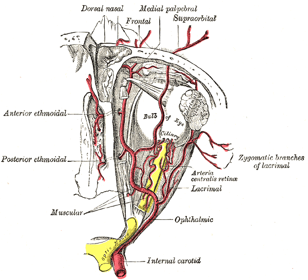
Three arteries carry the bulk of this risk because each links toward the eye. The supratrochlear and the supraorbital arteries climb the forehead from the inner brow. Both are branches of the ophthalmic artery itself. The dorsal nasal artery runs on the bridge of the nose and connects to the same ophthalmic supply. A bolus in any of the three has a short route straight back to the eye.
The events are rare. They are also the worst outcome in aesthetic medicine. A single one can follow a routine treatment. This is why an injector reads the arteries of the face before the needle. The few that connect to the eye get the closest attention. The map is a way to keep the needle out of those arteries in the patient on the table, whose vessels do not run exactly where a textbook draws them.
The glabella, between the brows
The ophthalmic artery, a branch of the internal carotid, and the vessels it gives off in the orbit: the supraorbital and frontal (supratrochlear) arteries that climb to the forehead, the dorsal nasal that runs to the nose, the central retinal artery to the eye. Filler forced back along the forehead or nose branches can reach this supply and the retina. This is an anatomy engraving (Gray’s Anatomy). It is not an ultrasound image. The red vessels, the yellow optic nerve and the labels are part of the original.
The glabella is the riskiest area on the face for filler. Two small arteries sit here: the supratrochlear, near the midline above the inner brow, and the supraorbital, a little further out along the upper rim of the orbit. Both come straight off the ophthalmic artery. Both run from the deep tissue up toward the skin as they climb.
What makes the glabella so unforgiving is the lack of spare supply. These arteries have little collateral circulation, so a vessel blocked here has no neighbor to take over. The skin it fed dies. The same vessel offers the filler a short path back to the eye. A scan here finds the supratrochlear and the supraorbital, reads their depth, and shows the plane an injection can use without crossing them.
Knowing the depth matters as much as the side-to-side position. These vessels sit deep near the brow and rise toward the surface higher up. A filler placed in the wrong plane meets the artery where a careful one would have passed above or below it. The screen turns the plan from a guess about an average glabella into a placement read off this one. The supratrochlear in particular dives deep at the brow and climbs to just under the skin a few centimeters up the forehead, so the same artery sits at two different depths within one field, deep low down and shallow higher up.
The nose
The nose carries more reported harm than any other filler site. Reviews put it as the leading cause of skin death from filler and the second leading cause of vision loss. The reason sits in its blood supply. The dorsal nasal artery runs down the bridge, fed from the ophthalmic system, with a direct path back to the eye. The lateral nasal arteries run along the sides near the tip. The angular artery climbs the side of the nose from the cheek.
These vessels run close to the surface on a structure with little soft tissue to spare. A filler meant to straighten a bridge or lift a tip sits in the same shallow layer the arteries use. The nose also draws filler for non-surgical reshaping more often each year, which puts more needles into this crowded supply. The skin of the nose is thin and the vessels sit just beneath it, so a plane that would be safe on a cheek runs dangerously shallow here. A small bolus goes a long way on a nose. The same small bolus reaches an artery sooner.
A scan before a nose treatment maps the dorsal and lateral vessels and the angular as it arrives. The injector reads where they cross the planned line and at what depth. Many injectors move to the deep, pre-periosteal plane against the bone here, where the vessels are fewer. The scan confirms the needle is in it. A blunt cannula in place of a sharp needle adds another margin. The screen shows the cannula staying clear.
The tip and the columella, the strip between the nostrils, carry their own small vessels from the lateral nasal and the columellar branches. These are easy to catch in a tip-refining injection. A scan of the tip before the needle shows how shallow the supply runs there. The same caution covers a surgical rhinoplasty, where a blade follows the same vessels a needle would.
The nasolabial fold and the cheek
The fold that runs from the nose to the corner of the mouth sits over the facial artery. The artery climbs from the jaw, crosses near the corner of the mouth, and runs up toward the nose, where it becomes the angular artery. Its exact path wanders from one face to the next. It can run just under the skin in the fold or deeper against the muscle. Studies of the facial artery find it taking several named paths through the midface, so no single rule places it for every patient. The injector who maps it reads the path this face took, not the one a diagram drew.
The depth decides the safe plane. In the lower two thirds of the fold, the injector has room in the dermis and the layer just below it. The upper third, near the nose, is tighter. A common rule places filler there in the deep pre-periosteal plane, a few millimeters above the groove of the nostril, to stay off the facial artery and vein. A scan reads which plane is clear in this patient and where the artery sits closest to the surface.
The cheek shares the same supply higher up. The angular artery and the branches that feed the mid-face run through it. A bolus in the medial cheek can reach the angular. The angular connects on toward the eye. The scan carries the same value here as at the nose: it shows the vessel before the needle finds it.
The lips
The lips run on the superior and inferior labial arteries. These loop through the body of each lip, usually between the muscle and the inner lining, a few millimeters in from the red margin. Their exact depth and path vary, which is why a lip can bleed or bruise from a needle that found the vessel by chance.
A lip bruises more than it threatens.
The labial arteries carry less of the blindness risk than the glabella or the nose. No clear case of blindness has come from a lip treatment. The superior labial artery does connect upward through the face, so a theoretical path exists. The everyday gain from a scan here is steadier: fewer vessels caught, less bruising, and a clearer sense of the plane to fill. A scan shows the loop of the artery so the needle can sit shallow to it or deep to it. The gain on the lips is steadier than dramatic. A scan that finds the labial loop spares the patient a bruise and the clinician a callback, even where the blindness risk is remote.
The forehead and the temple
The forehead continues the glabella’s supply upward. The supratrochlear and supraorbital arteries fan out across it from the inner brow, climbing from deep to shallow as they rise. Filler smoothing a forehead line sits in the layer these vessels pass through on their way up. A scan reads their depth at the line being treated. The danger on the forehead is the glabella’s, carried upward. A bolus forced into a forehead branch can travel back down to the eye along the same vessel it climbed. The high forehead feels far from the eye to the hand. It is not far along the artery.
The temple holds its own hazard. The superficial temporal artery runs across it under the skin. A large vein, the sentinel vein, sits in the same area. A needle into the vein can send filler toward the deep veins behind it. A needle into the artery can carry filler toward connections with the eye’s supply. The temple also has a deep plane right against the bone. A scan confirms the needle reaching it past the vessels above.
Veins carry a risk too
The arteries carry the gravest risk. The veins carry one of their own. Filler pushed into a facial vein can travel with the venous flow toward the deep veins of the head. The angular vein near the nose connects back toward the cavernous sinus behind the eye. A clot or a plug of filler reaching there brings its own emergency. The same scan that maps the arteries shows the larger veins, so the needle can stay clear of both.
Color Doppler tells a vein from an artery on the same scan. A vein shows a steady low flow and flattens under light pressure from the probe. An artery pulses and holds its shape. The injector reads both as the probe sweeps, so the map covers the whole vascular field, the veins included. A vein and an artery can show the same color, since the color marks direction, not vessel type. A blue vessel is not always a vein. A red one is not always an artery. The injector tells them apart by the pulse and the squeeze of the probe, not by the hue. Mistaking the two matters, because the arteries are the ones that carry the danger back toward the eye.
Putting the map to work
The map is only the first half. The injector reads it into a plan before the needle moves. The plane the filler will sit in gets chosen against the depth of the vessels, so the product lands where the mapped arteries do not run. The entry point and the angle follow from the same picture. A vessel crossing the planned path turns the plan toward a different entry or a deeper plane.
The map also decides the tool. Where the arteries run shallow and crowd together, as on the nose and the glabella, the injector reaches for a blunt cannula in place of a sharp needle, since a cannula pushes vessels aside where a needle would pierce them. The map shows whether there is room for a cannula to pass without crossing an artery. It shows whether the plane is deep enough to trust, or so crowded that the safest move is to place less, or nothing at all, in that spot. A high-risk area with a vessel running straight through the planned site is a reason to change the plan, not to work around a known danger. The injector marks the vessels on the skin or keeps the probe in hand through the injection, so the picture stays present while the needle moves. A map read once and then forgotten loses its value the moment the injector looks away. The whole point is to carry the picture all the way to the needle. Admiring it and then injecting from memory misses the point.
The same picture guides the rescue if it is ever needed. An injector who mapped the arteries going in knows where they run if a vessel blocks. The probe that found the artery is the fastest way to find the trapped filler afterward. That rescue is a subject of its own. For the map, the point is narrower: the scan done before the needle pays off twice.
Why a map beats a remembered diagram
A textbook shows the average face. The arteries on a real face run a centimeter off that average often enough to matter. The facial artery can cross the fold high or low. The dorsal nasal can run more to one side. The supratrochlear can sit deeper than the diagram suggests. A memorized map gives a starting point. It does not give this patient’s anatomy.
The scan closes that gap. Color Doppler shows where each artery runs in the face on the table, at what depth, and where it sits closest to the planned injection. The injector plans the entry and the plane against that picture. The diagram tells the clinician which arteries to look for. The scan tells the clinician where they are this time.
What the list does not cover
The map shows the arteries the probe can resolve. The smallest vessels run below what even a high-frequency probe sees, so a clear scan lowers the risk of an intravascular injection. It does not erase it. The list of danger arteries is a guide to where to look. It is not a promise that a found path is the only one. A careful injector reads the scan as one strong safeguard among several, alongside slow injection, a blunt cannula in the riskier areas, and a plan for treating an occlusion if one happens. The list is a guide to where to look hardest, drawn from the cases that have caused harm over the years. A vessel can still surprise an injector who treats the map as the last word. The safe habit is to scan every high-risk site fresh on the day, then trust the screen over the memory of an average face.
Common questions
Which facial arteries carry the risk of blindness?
The ones that connect back to the ophthalmic artery behind the eye. The supratrochlear and supraorbital arteries on the forehead and the dorsal nasal artery on the nose are the main ones, since each links to the ophthalmic supply. Filler forced backward through any of them can reach the vessels of the retina.
Why is the glabella the riskiest area?
It holds the supratrochlear and supraorbital arteries, both branches of the ophthalmic artery, with little spare supply around them. A vessel blocked here has no neighbor to take over, so the skin dies. The same vessel offers a short path back to the eye. A scan finds these arteries and the safe plane before the needle.
Why does the nose cause so many complications?
Its arteries run shallow on a structure with little soft tissue. The dorsal nasal artery has a direct route back to the eye. Reviews list the nose as the leading cause of skin death from filler and the second leading cause of vision loss. A scan maps these vessels and confirms the needle stays in the deep plane against the cartilage.
Are the lips a high-risk area?
Less so than the glabella or the nose. No clear case of blindness has come from a lip treatment. The labial arteries still bleed and bruise when a needle finds them. A theoretical path to the eye exists through the superior labial artery. A scan shows the loop of the artery in each lip so the needle can sit clear of it.
Does a textbook diagram replace a scan?
No. A diagram shows the average face and tells the injector which arteries to look for. The arteries on a real face run off the average often enough to matter. A scan shows where they run in this patient, at what depth, so the plan fits the face on the table.
Does ultrasound show the veins as well as the arteries?
Yes. Color Doppler shows both. A vein carries a steady low flow and flattens under the probe. An artery pulses and holds its shape. The veins matter because filler in the angular vein near the nose can travel back toward the cavernous sinus behind the eye. A full map covers both.
Does mapping the arteries remove the risk entirely?
No. The smallest vessels run below the probe’s resolution. Anatomy varies. Mapping lowers the chance of an intravascular injection by a large margin. A careful injector treats it as one safeguard among several, alongside slow injection, a blunt cannula in the riskier areas, and a plan to treat an occlusion if it happens.
About the Author
Julien Mercier
Senior R&D Engineer · Medical Ultrasound Transducer Development
Senior R&D Engineer with an M.S. in Applied Physics and over 15 years of experience in medical ultrasound transducer development, specializing in the design verification and performance testing of high-frequency imaging transducers. Currently leading the development and verification of the company’s next-generation high-frequency linear-array transducer, responsible for imaging performance evaluation and reliability analysis in preclinical testing. Brings extensive hands-on experience in piezoelectric element tuning, beamforming parameter optimization, and system-level performance testing.