Ultrasound localizes the muscle a botulinum toxin injection is meant for. The injector sees the target muscle on the screen and watches the needle enter it. Blind landmarks miss that muscle more often than injectors expect.
Why a muscle is hard to hit blind
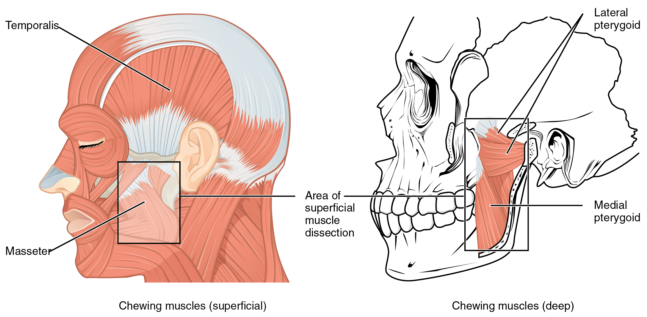
The chewing muscles, with the masseter at the jaw, a frequent target for botulinum toxin. The probe finds it by sight before the needle goes in. This is an anatomical diagram. It is not an ultrasound image. The labels are part of the original.
Botulinum toxin works on the muscle it reaches. The dose has to land in the right muscle, at the right depth, in the right part of that muscle. A landmark technique aims by surface marks and feel. It assumes the muscle sits where an average face would put it. Faces vary. A muscle can sit deeper, run wider, or end sooner than the marks predict.
One masseter study put numbers on this. Surface marking placed the injection outside the target muscle in up to four cases in ten. The needle sat in fat or in a neighbor, away from the muscle the dose was meant for. The injector could not tell. The mark looked right from the surface. The toxin went where the needle went. The misses were silent. The injector felt the needle reach what seemed the right spot, with no way to know it sat in fat or in a neighbor. Only the scan shows the gap between where the needle felt right and where it landed.
The depth matters as much as the spot on the skin. A muscle a centimeter down takes a different needle angle than one just under the dermis. A dose placed too shallow spreads in the fat above the muscle. A dose placed too deep reaches a muscle below the target. Surface feel gives a rough depth at best. The screen gives the real one.
The masseter makes the depth problem concrete. It runs in two layers, a superficial part and a deep one. The two can take different amounts of toxin. A needle set for the surface reaches only the superficial layer. The deep part keeps working. The jaw stays wide on that side. Reading the full thickness of the muscle on the screen lets the dose reach the layer that needs it.
Seeing the muscle on the screen
On ultrasound a muscle has a look of its own. It shows as a band of tissue with fine internal lines, bounded by brighter sheets of fascia above and below. The probe runs over the area, and the target muscle stands out from the fat and the muscles around it. The injector reads its width, its depth, and the edge where it ends.
The picture is live. The scan can move. The clinician presses gently and watches the muscle shift, or asks the patient to clench and sees it thicken. A muscle that contracts on cue confirms itself. The clench also shows how far the muscle reaches and where it sits deepest. The needle then enters under the same live view. It shows as a bright point pushing into the band. The dose can be placed in the belly of the muscle, away from its edge.
The thickness becomes a number to work from. A strong masseter in someone who clenches can measure a centimeter or more across. A slack one measures far less. That number sets how much toxin to place and in how many spots. A thick muscle takes more, spread across the bulk. A thin one takes less. The dose then follows this muscle’s own number.
Putting these readings together turns the scan into a plan before the needle moves. The injector first finds the muscle and traces its outline, noting where it begins and ends so the dose stays inside it. A clench then shows the working part of the muscle, the bulk that pulls hardest, the part a dose is meant to weaken. The thickness measured at the deepest point sets the amount and the depth. A thin muscle treated like a thick one ends up over-dosed. A thick one treated like a thin one barely moves. The injector also notes what sits around the muscle, a gland behind it, a vessel along its edge, a neighbor a few millimeters off, so the needle has a path that reaches the target and clears the rest. By the time the needle goes in, the injector is not guessing at any of it. The whole injection has been rehearsed on the screen. The needle only carries out what the picture already settled.
The masseter, the prime example
The masseter is the clearest case for the technique. It is the muscle treated for a wide jaw and for clenching at night. It varies in size from one face to the next, which is the whole reason a dose needs placing by sight. A large masseter takes more toxin across more of the muscle. A thin one takes less, in fewer spots.
The masseter also sits in a crowded corner. Its back edge runs up against the parotid gland, the largest salivary gland. A needle that drifts back from the muscle can reach the gland. Salivary tissue varies too, with one study finding a variant in roughly two thirds of people. Ultrasound shows the back edge of the muscle and the gland behind it. The dose stays in the muscle.
A strong masseter takes its dose spread across the bulk. The clinician places several small amounts, each one inside the muscle on the screen, so the whole muscle relaxes evenly. A single large bolus in one corner leaves the rest of the muscle working. The result looks lumpy or uneven. Watching each placement keeps the dose spread the way the plan intends.
The scan helps again at the follow-up visit. A jaw that stayed wide can be checked for a deep layer the first dose never reached. A masseter that thinned on one side shows an uneven first treatment. The next dose then goes where the first one fell short. The screen turns a touch-up from a guess into a correction aimed at a known spot.
The scan of a masseter is quick to run. The probe sits flat on the cheek over the angle of the jaw. A clench brings the muscle up under it. The injector reads the thickness, marks the safe spread, and places the needle in-plane so its track shows on the screen. The whole check adds about a minute to a treatment the clinic already does. That minute is the cheapest part of the visit.
The crowded face and the wrong muscle
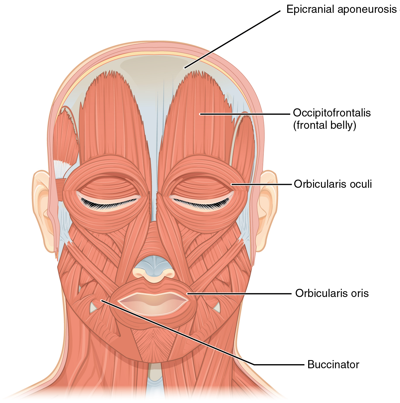
The muscles of facial expression, frontal view: the frontalis that raises the brow, the orbicularis around the eye and the mouth, the platysma down the neck. These are the small muscles a botulinum dose has to reach without weakening a neighbor. This is an anatomical diagram. It is not an ultrasound image. The labels are part of the original.
Faces run on small muscles packed close together. A few millimeters separate one from the next. The muscle that lifts the lip sits beside the one that flares the nostril. The muscle that pulls the mouth sideways sits below the cheek. A dose meant for one muscle weakens its neighbor when it lands a little off.
A few millimeters decide which muscle takes the dose.
The errors are specific, and each one shows on the face. A dose meant for the frontalis, set too low, drops the brow and leaves the lid heavy. A dose near the eye that spreads to the wrong fibers leaves an eyelid that droops for weeks. A dose in the neck that reaches the muscles of swallowing makes swallowing hard. Each follows a needle placed a little off in a crowded field.
These complications fade as the toxin wears off, over weeks to months. The wait is the patient’s to bear. A crooked smile or a heavy brow on a face the patient came in to improve is its own kind of harm. Seeing the target muscle and its neighbors on the screen lowers the chance of that wait. The dose goes into the intended muscle the first time. The cost of the miss is visible and lasting, weeks of a crooked smile or a heavy brow on a face the patient paid to improve. The scan trades a minute of looking for that risk.
Placing the dose
Seeing the muscle is half the job. Placing the dose well is the other half. The toxin spreads a short distance from where it lands, so the depth and the spot decide which fibers it reaches. A dose in the belly of a thick muscle stays in that muscle. A dose near an edge can drift into a neighbor as it spreads. The screen shows the edge, so the placement keeps its distance from it. The spread is the reason depth matters so much. A dose sitting a few millimeters too shallow can creep up into the muscle that lifts instead of the one that clenches. The screen lets the injector place the tip where the spread stays inside the target. The spread also sets a floor on the dose. Too little in a thick muscle leaves part of it working. The result looks half-treated. The screen shows the bulk to be covered, so the amount matches the muscle in front of the clinician.
Keeping the needle visible is part of the placement. The beam is a thin plane. The needle shows in full only while it stays inside that plane. The clinician lines the needle up with the beam and watches the tip reach the chosen depth inside the muscle. A tip lost outside the plane is a dose placed blind, even with the muscle in clear view.
The live needle makes the whole injection deliberate. A large muscle can take several small doses across it, each one checked on the screen, so the effect comes out even. A muscle close to a vessel or a gland lets the clinician steer the tip away before pressing the plunger. The injection becomes a thing the clinician can see on the screen. The mind no longer pictures the path.
Reading the muscle before and after
The scan has a place at both ends of the treatment. Before the needle, it maps the muscle, its layers, and the structures around it. The clinician sets the depth, the points, and the dose against a real picture of this patient. A measured thickness gives a baseline. The whole plan rests on what the muscle is. A photo of the screen, saved with the notes, fixes that baseline in the record. The next visit opens it and measures against it rather than against memory.
Weeks afterward the scan reads the result. A treated masseter thins as the toxin takes hold. The screen shows by how much. A muscle that barely changed points to a dose that missed the target or fell short. The first scan sits beside the new one, so the comparison is a measurement. The next plan builds on what the muscle did, which is more than a patient’s report or a glance in the mirror can give. The number also settles a dispute. A patient who feels the treatment did nothing can be shown a muscle that thinned on the scan, or an honest reading that it did not. The screen replaces an argument with a measurement.
Other muscles around the face
The masseter is the busiest target. The same approach reaches the smaller muscles of the upper and middle face. The frontalis raises the brow. A dose set too low on it drops the brow. The depressor that pulls the corner of the mouth down sits close to the muscles that move the lip. The mentalis bunches the chin. Each is a thin sheet a few millimeters down, packed against neighbors that do the opposite job. A scan shows the depth of each one and the gap to the next muscle.
The value rises with the risk of the area. Around the eye and the mouth, a dose that strays gives a droop or a crooked line the patient sees every day for weeks. The thin muscles there reward a careful depth and a placement set back from the neighbor. Ultrasound gives both. The broad muscles of the forehead carry a wider margin. Many injectors treat those by feel. The case for the scan grows as the muscle gets smaller and the neighbors sit closer.
Beyond the face
The same approach guides toxin into muscles past the jaw and the brow. The platysma runs from the collarbone up to the jaw. Toxin along its upper edge softens the bands that stand out when the neck tenses and sharpens the line of the jaw. A scan finds that edge and the depth the muscle sits at. The dose stays clear of the structures lower in the neck. The platysma is a thin sheet, easy to inject too deep, where the toxin would reach the muscles that move the voice box. The scan keeps the needle in the sheet.
A salivary gland moves the target from a muscle to a gland. Toxin placed into the gland eases heavy drooling, a problem in some neurological conditions. The gland has to be seen to be dosed, since vessels and nerves run close around it. The frontalis, the corrugators between the brows, and the muscles around the mouth each have the same theme. A small muscle sits near something the dose should avoid. The screen shows the way around it. Each of these targets sits near something the dose must spare: a nerve, a gland, a vessel. The scan turns a risky blind injection into a placed one.
When the muscle is hard to see
Some muscles read cleanly. Others fight the probe. A heavy layer of fat over the cheek pushes the masseter deeper and dims the picture, so the injector turns up the depth and accepts a softer image. A thin face brings the muscles close to the skin, where they crowd together and the gap between two of them shrinks to a millimeter. Each case asks the injector to tune the machine to the face in front of them. A standard setting fits no one in particular.
A muscle treated before reads differently. Toxin given months earlier leaves a muscle thinner than its mate on the other side. Old scar from a deep injection breaks up the clean fibers the probe looks for. The injector compares the two sides, since a muscle that should match its partner and does not tells its own story. The history of the face shapes what the screen shows.
The smallest target muscles test the probe’s limits. A muscle the width of a few millimeters, lying under another, can be hard to separate on the screen. The injector leans on the clench here, since a muscle that thickens on cue marks itself out from the still tissue around it. Where even that fails, the dose stays conservative and the injector treats the area as the crowded place it is.
What it does not do
Ultrasound does not choose the dose or the plan. A clinician still decides which muscle to treat and how many units to place. The probe does not steady a careless hand. It does not work without an operator who can read the muscle from the fat around it. The skill of reading the picture comes with practice, over a few dozen scans. The probe shows the target. The clinician still has to hit it.
Common questions
Why use ultrasound for a botox injection?
It shows the target muscle directly. A landmark technique aims by surface marks. One masseter study found those marks placed the needle outside the muscle in up to four cases in ten. Ultrasound puts the dose in the intended muscle and keeps it off the nerves, glands and neighboring muscles nearby.
Which botox treatments gain first from it?
The ones where the muscle is hard to find by feel or sits near something delicate. The masseter is the clearest, since it varies in size and runs against the parotid gland. The platysma in the neck and the salivary glands for drooling also gain, because each sits near a structure the dose should avoid.
Can a misplaced dose cause harm?
It can cause a visible problem. A dose that drifts to the wrong muscle can pull a smile crooked, drop a brow, or weaken a muscle the patient needs. The effect fades as the toxin wears off over weeks to months. Seeing the muscle and the needle lowers the chance of it.
Is the masseter that close to the salivary gland?
Yes. The back of the masseter runs against the parotid gland, the largest salivary gland. A needle that drifts off the muscle can reach the gland. Ultrasound shows the back edge of the muscle and the gland behind it, so the dose stays where it belongs.
Can ultrasound show whether the treatment worked?
Yes, by comparing scans. A treated muscle thins over the weeks after a dose. Measuring its thickness before and again later shows the change as a number. A muscle that barely thinned points to a dose that missed or fell short. The next session can then correct it.
Does it help with the deeper part of the masseter?
That is one of its main uses. The masseter has a superficial and a deep layer. A dose set for the surface can leave the deep layer working, so the jaw stays wide. Reading the full thickness on the screen lets the clinician reach the layer the surface dose missed.
How hard is the technique to learn?
Reading a muscle from the fat around it takes practice, on the order of a few dozen scans. Holding the probe still and keeping the needle in the plane of the beam are the basic skills. The harder part is turning the scan into a routine step. Reaching for it only when a case looks difficult misses the point.
About the Author
Julien Mercier
Senior R&D Engineer · Medical Ultrasound Transducer Development
Senior R&D Engineer with an M.S. in Applied Physics and over 15 years of experience in medical ultrasound transducer development, specializing in the design verification and performance testing of high-frequency imaging transducers. Currently leading the development and verification of the company’s next-generation high-frequency linear-array transducer, responsible for imaging performance evaluation and reliability analysis in preclinical testing. Brings extensive hands-on experience in piezoelectric element tuning, beamforming parameter optimization, and system-level performance testing.