Ultrasound Facial Vascular Mapping Before Hyaluronic Acid Filler Injection
Vascular mapping before a filler injection is the scan an injector runs to find the arteries in a face before the needle does. It is done with color Doppler on a high-frequency probe, swept in a set pattern over the area about to be treated. Done well, it turns the riskiest guess in the procedure into something the injector can see on a screen.
Setting up the scan
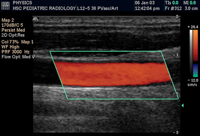
A color Doppler image: the vessel fills with the color of its flow inside the sampling box. The scale, marked PRF at the top, is the setting that decides which flows show. A facial map is built from the same signal, set for the slow flow of a small vessel. This is a Doppler example on a peripheral artery. It is not a facial scan. The labels are the machine’s own settings.
The scan starts with the settings, because a facial artery is small and its flow is easy to miss. The probe runs at the high end, around 18 MHz or above, for the resolution to show a vessel a millimeter or two across. Color Doppler does the main work. It fills a moving vessel with color, so an artery that is invisible on the plain gray image appears the moment blood runs through it. The picture updates live on a phone or a tablet screen while the probe moves.
The one setting that decides everything is how slow a flow the machine will show. A facial vessel carries slow flow compared with the arteries in a limb. If the scale is left where it sits for a wrist or a neck, the probe runs over a real facial artery and shows nothing. The injector then reads an empty screen as a clear path. This is the worst failure of mapping, because it looks exactly like success. The scale has to be set low, for slow flow at a shallow depth, before the scan means anything.
Power Doppler is the backup for the smallest, slowest vessels. It is more sensitive than color Doppler, with no direction shown. Spectral Doppler is the tie-breaker. Placed on a single vessel, it reads out the shape of the flow over time, which settles any doubt about what the vessel is. A map is usually made on color alone, with power and spectral reached for when a vessel is faint or unclear.
A clinician confirms the settings before trusting them. The simplest check is to find a vessel known to be there, the facial artery at the jaw, and see that it lights up. A machine that shows a known artery is set to show the unknown ones. A quiet screen over a vessel that should be loud is a setting problem. It is not a safe area. Treating it as safe is how the scan fails silently.
The sweep
Mapping is a routine. It is not a glance. The injector sweeps the probe across the whole area about to be treated, slowly enough for the color to fill each vessel as the beam crosses it. The sweep is run in two directions, because a vessel that runs along the beam can slip past a single pass. The aim is a mental picture of where every artery in the field runs, built from a flat beam moved through a network that sits at several depths.
The sweep follows a discipline, not an impulse. The injector starts at one edge of the field and moves the probe in slow overlapping passes, so no strip of skin goes unread between two sweeps. Each pass runs slowly enough that the color has time to fill a vessel before the beam moves on, since a fast pass paints a slow artery the same gray as the tissue around it. The probe then crosses the field again at a right angle to the first set of passes, because an artery running straight along the beam can hide in one direction and only show when the beam meets it across its width. Through all of it the injector watches the depth scale at the side of the screen, building a picture that holds not only where each vessel runs but how deep it sits at each point. By the end the injector carries a three-dimensional sense of the field, the arteries and their depths together, rather than a flat tracing of lines on a surface. That picture is what the needle gets planned against.
The high-risk regions get the closest attention. Those areas sit over the midface, where arteries connect back toward the eye and a blocked vessel does the gravest harm. In each region the scan follows the known course of the artery and marks where it runs in this face, often a centimeter off the textbook line. The exact vessels that matter in each area are a study of their own; the sweep is how an injector finds them in the patient in front of them.
Depth is read along with position. A vessel that sits deep to the plane a filler will fill is a different risk from one that crosses that plane directly. The injector notes where each artery runs across the face and how deep it sits, because the same vessel can be safe at one level and squarely in the needle’s path at another. A map that records position without depth is only half a map. The depth scale at the side of the screen carries that second number for every vessel the sweep crosses.
Telling an artery from a vein
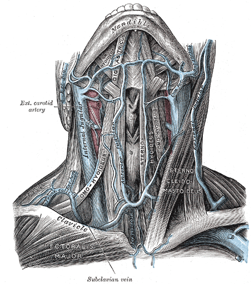
The arteries and veins of the head and neck: the external carotid here gives rise to the facial artery, with the jugular veins alongside. A scan tells the two apart by pulse and compressibility. Color does not decide it. This is an anatomy engraving. It is not an ultrasound image. The labels are part of the original.
Color alone does not say which vessel is an artery. The color shows only direction: blue for flow heading away from the probe, red for flow heading toward it. A blue vessel is not a vein. A red one is not an artery. The mistake of reading color as vessel type is common. It is also dangerous, because the whole point of the scan is to respect the arteries.
Three other signs settle it. The first is pulsation: the beat of the heart runs visibly through an artery. A vein holds a steadier flow. The second is compressibility. A vein flattens under the light pressure of the probe. An artery holds its shape against the same push. The third is velocity, which the scale on the screen reads out: an artery runs faster than the slow flow of a vein. Any one sign can mislead. Together they are reliable.
The discrimination matters because the consequences split on it. An artery, injected, can block blood to the skin or the eye. A vein taking filler is a lesser event, uncomfortable and usually without lasting harm. So the effort goes into finding and respecting the arteries above all. When a vessel cannot be classified with confidence, the safe reading is to treat it as an artery and stay clear. Confidence comes with repetition. Early on a clinician confirms each vessel with the spectral waveform. With practice the pulse and the compressibility carry nearly every reading at a glance.
Depth, not just position
A face is not a flat map. The arteries weave between the skin, the fat compartments and the muscle, changing depth as they go. A vessel that runs deep in one spot surfaces toward the dermis a centimeter on, which is exactly where a superficial filler would meet it. Mapping in two dimensions, marking only the line on the surface, misses this. The injector reads each vessel as a path in three dimensions.
This is why the plane the filler will sit in is part of the plan. An injector decides the depth of the injection against the depth of the vessels, choosing a plane that the mapped arteries do not cross. Where a vessel and the intended plane meet, the plan changes: a different entry, a different depth, or a blunt cannula in place of a sharp needle. The scan turns the depth of the injection from a feel into a decision made against a picture. The same depth read decides the layer for the product. A filler meant for the deep fat sits below the bulk of the vessels. One placed in the superficial skin runs closer to them. The scan tells the injector which planes are clear at the chosen spot before the needle commits to one.
From the map to the needle
Mapping is half the technique. The other half is keeping the picture live while the filler goes in. One described approach uses three steps: map the arteries first, inject under real-time ultrasound, then check that blood is still flowing afterward. A retrospective report followed this approach in around 480 patients as a way to lower vascular events in high-risk areas.
In practice the injector holds the probe in one hand and the syringe in the other. The needle tip shows as a bright point. The filler appears as a dark pocket spreading from it. The injector watches both against the mapped vessels, so the product lands in the planned plane and stays clear of the arteries. A bolus drifting toward a vessel shows on the screen with time to stop, which is the difference real-time guidance makes over working from a map drawn and then set aside.
Keeping the needle useful on screen is its own skill. The beam is a thin plane, so the whole shaft of the needle shows only when it stays inside that plane. An injector learns to line the needle up with the beam, so the screen shows the shaft as a full bright line. A single dot on screen could be the tip or any point along the shaft. Lose the needle in the beam and the guidance is gone, even with the vessels in clear view.
The map also pays off if the worst happens. If a vessel is hit, the same probe finds the deposit and guides the enzyme that dissolves it. That rescue is a subject of its own. For mapping, the point is narrower: the scan that found the artery before the needle is also the fastest way to find it afterward. A clinic that mapped going in already knows the local anatomy when seconds count.
None of this works as a one-time lesson. The hand learns the probe. The eye learns the flow. Bringing the two together while a needle moves takes longer than either alone. It is a skill built over many scans. The injectors who trust it have done it on the faces where nothing went wrong, not only on the one where something did.
When the map is hard to read
Some faces fight the scan. Old filler from an earlier treatment scatters the sound and throws shadows that hide a vessel behind them. A hyaluronic acid deposit reads as a dark pocket that can sit right where an artery runs, so the injector has to work the probe around it from more than one angle. Scar tissue from surgery or from a previous thread does the same, breaking up the clean planes the beam needs. The map in these faces takes longer and leans harder on the spectral waveform to confirm what a cluttered gray image cannot.
Swelling changes the picture too. A face injected days earlier, or one carrying fluid for another reason, holds tissue that looks different from the textbook layers. Vessels sit deeper than usual under the extra fluid. The injector reads the depth fresh rather than trusting a number from a thinner face. A scan repeated once the swelling settles often reads more cleanly than one rushed while the tissue is still puffy.
The hardest vessels are the smallest. A tiny branch carrying a trickle of flow can sit below what the probe resolves, showing as nothing on color and little on power. The injector treats the area around a known larger artery as risky whether or not a small branch lights up, since absence of color over a small vessel is the one reading the scan cannot fully trust. The map marks the vessels it can prove and flags the zones where an unseen branch is likely.
Holding on to the map
A map is only useful while it is in mind. Some injectors draw the vessels they found on the skin before they pick up the needle, turning the scan into marks they can see while they inject. Others keep the probe in hand and treat under live imaging, so the map never has to be remembered at all. Either way, the injection follows a path checked in this patient minutes before.
The scan can also be saved. An image of the mapped vessels, stored with the treatment notes, records that the area was checked and shows the anatomy for the next visit. A map made and then forgotten helps little, because filler can be pushed along a plane and reach a vessel the scan had shown earlier. The discipline of mapping is as much about holding the picture through the injection as about making it in the first place.
Where it goes wrong
The commonest failure is the settings, already named: a scale left too high, so a real artery reads as a quiet area that looks safe. It is the gravest error, because it rewards carelessness with a clean-looking screen. The guard against it is the habit of setting the machine for slow flow and confirming on a known vessel every time.
A high scale hides the one thing the scan is for.
The next failure is the rushed sweep. A probe moved too fast, or in one direction only, skips vessels that a slower, two-way sweep would have caught. Mapping cannot be hurried into a few seconds and still be trusted. The injector who treats it as a box to tick gains little from owning the probe.
The last failure is overconfidence. A clinician who reads a fast map without the checks, or who trusts a two-dimensional line and forgets depth, can be more dangerous than one who never scanned, because the scan lends a false certainty. The map is a tool for a careful hand. It does not replace the care. Used as a substitute for it, it can mislead.
What mapping does not promise
Mapping lowers risk; it does not erase it. The smallest vessels can run below what even a high-frequency probe resolves. Anatomy varies. A vessel can sit just outside the swept area. The scan is only as good as the settings and the hand behind it. A clean map is a strong reason for confidence. It is not a guarantee. The careful injector reads it that way.
The map also goes stale as the face changes. Filler placed in one session shifts the tissue for the next. A map drawn months ago describes a face that no longer exists. Each treatment starts with a fresh scan rather than a saved one. The stored image guides the next visit as a starting point, a reminder of where the arteries ran. It is not a substitute for looking again.
Common questions about vascular mapping
What ultrasound mode is used to map facial vessels?
Color Doppler does the main work, filling a vessel with color when blood moves through it. Power Doppler is added for the smallest, slowest vessels, since it is more sensitive. Spectral Doppler, placed on one vessel, reads its waveform to confirm what it is. All three run on a high-frequency probe set for slow flow at a shallow depth.
How do you tell an artery from a vein on the scan?
Not by color, which only shows direction. Three signs do it. An artery pulses with the heartbeat. It resists the light pressure that flattens a vein. Its velocity on the scale runs higher than a vein’s slow flow. When a vessel cannot be classified for sure, it is treated as an artery and avoided.
What settings make the vessels show on the scan?
Color Doppler, set for the slow flow of small facial vessels. The pulse repetition setting comes down so the scan catches low velocities. The gain comes up until the artery fills with color. Too much gain floods the whole screen. A light touch on the probe matters too, since pressing flattens a vein and hides it.
Does mapping mean the injection is guided live as well?
Usually, yes. The strongest technique maps the vessels, injects under real-time ultrasound, then checks that flow continues afterward. The injector watches the needle and the filler spread against the mapped arteries, so a bolus heading the wrong way shows in time to stop.
Can a scan miss a vessel?
Yes. The smallest vessels can run below the probe’s resolution, anatomy varies, and a vessel can lie just outside the area swept. A scale set too high can also hide a real artery. Mapping lowers the risk of an intravascular injection a great deal. It does not remove it.
Does old filler make the scan harder?
Yes. A hyaluronic acid deposit reads as a dark pocket and scatters the sound, throwing shadows that can hide a vessel behind it. The injector works the probe around the deposit from more than one angle and leans on the spectral waveform to confirm what the gray image cannot. The map in a face full of old filler takes longer.
Which one setting decides the scan?
The one that controls how slow a flow the machine will show, often called the scale or the PRF. Set for a limb artery, it makes a slow facial vessel disappear. Set low, for slow flow at a shallow depth, it shows the vessel. Confirming the setting on a known artery before each scan is the simplest safeguard.
About the Author
Julien Mercier
Senior R&D Engineer · Medical Ultrasound Transducer Development
Senior R&D Engineer with an M.S. in Applied Physics and over 15 years of experience in medical ultrasound transducer development, specializing in the design verification and performance testing of high-frequency imaging transducers. Currently leading the development and verification of the company’s next-generation high-frequency linear-array transducer, responsible for imaging performance evaluation and reliability analysis in preclinical testing. Brings extensive hands-on experience in piezoelectric element tuning, beamforming parameter optimization, and system-level performance testing.