Scarring, repeated over years, turns a soft liver into a cirrhotic one: hard, shrunken, its surface gone to bumps. Ultrasound catches that change and puts a grade on it, from the first faint coarsening to the hardened liver behind a wall of pressure. The grade is not academic. It sets how often a patient is watched for liver cancer, whether varices are hunted before they bleed, and where the liver sits on the path to failure or transplant.
The bumpy edge
Start at the surface, because the surface tells more than the rest. A healthy liver wears a smooth capsule, a clean bright line where it meets the wall in front of it. In a cirrhotic liver that line breaks into bumps. The scar and the regenerating tissue beneath push the edge up.
The bumps hide from the wrong probe. A deep convex probe blurs the surface into a soft haze. A high-frequency linear probe, laid over the front of the liver, brings the nodular edge into sharp relief, and where a little fluid has gathered, the ascites outlines the bumps like water lapping a bed of pebbles. The gain and the focus are set to the near field, where a deep abdominal preset would only blur the surface that holds the answer.
A surface gone frankly nodular is among the more specific things the scan can show. The reverse does not hold: a smooth-looking surface clears nothing, since the change arrives late and patchily. A clearly bumpy edge, in a patient with reason to scar, comes close to proof on its own.
The whole front of the liver is read for the surface, because the nodules come unevenly. A liver smooth in one place can be frankly bumpy in another, and the reader hunts for the worst of it. The probe is rocked along the edge, on a held breath, until the capsule shows plainly against the wall or the fluid beyond it.
A grain gone coarse
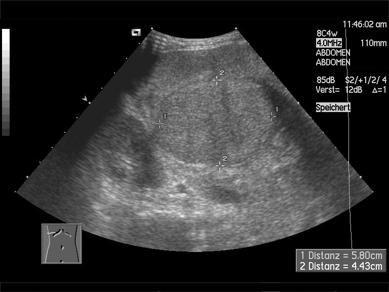
A cirrhotic liver on ultrasound. The smooth, even grain of a healthy liver has given way to a coarse, lumpy texture, the parenchyma broken up by scar and nodules. The calipers and the German labels are the machine’s own. Image: Christaras A, CC BY 2.5.
Run the beam through the liver itself and the texture has changed. A cirrhotic liver reads coarse, its even grain broken up by the nodules and the bands of scar that thread between them.
The coarseness is read with care, because a fatty liver coarsens too, and the two often sit together. Fat brightens the whole liver. As the beam travels deep, the fat dims it. Scar adds the lumpiness, the uneven stippling that fat alone does not bring. Telling the bright blur of fat from the coarse stipple of scar is part of grading the parenchyma. A heavily fatty liver also swallows the deep beam, so the far side of the liver and the diaphragm behind it fade from view. Turning up the power or dropping the frequency wins some of that depth back. The picture softens in return.
The regenerating nodules can sometimes be picked out as faint rounded shapes within the field. Many stay too small and too like their surroundings to see one by one. What the eye reads is their sum: a liver that has lost the smooth, uniform glow of health. A handful of larger regenerating nodules can stand out enough to be measured and followed, each one a question the reader keeps in view against the day it turns.
What scarred it
A grade reads better when the cause is known. Each cause leaves its own fingerprints. The scan is held beside the patient’s history, because the cause behind a coarse, bumpy liver changes what it means.
Viral hepatitis, B or C, scars the liver slowly from within over decades. The damage is diffuse. The organ it leaves is the classic shrunken, nodular liver the grading signs were drawn from. A patient with a long-carried virus is scanned with cirrhosis half expected. The same virus that scars the liver also drives its cancer, so these patients carry a doubled reason for the close watch a grade sets up.
Alcohol reaches the same end by a different road, often through a fatty, swollen liver that later shrinks and hardens. Early on the organ may be enlarged, its fat making it bright. Later it follows the same path to the small, knobbly block. The story across visits tells which stage the drinker has reached, better than any single scan can. A liver still swollen with fat may recover if the drink stops. Once it has shrunk and hardened, the point of return is past. The scan marks which side of that line a patient sits on.
Fatty liver disease, the kind tied to weight and the metabolism, has become the commonest road of all in many places. It begins as a bright, enlarged liver and can scar on to cirrhosis without a drop of drink. Its early brightness can mask the coarsening of fibrosis, one reason elastography counts for so much in these patients. The numbers of this disease are climbing with the weight of whole populations. The cirrhosis it leaves now fills clinics that once saw drink and virus alone.
Rarer causes leave their marks too: the blocked ducts of biliary disease, the iron-loaded liver of haemochromatosis, the autoimmune liver that turns on itself. Naming the cause leaves the grading signs unchanged. It still sharpens the reading of them, and it sets the company the liver keeps.
The liver redrawn
Cirrhosis does not shrink a liver evenly. It robs some parts and feeds others, and the lopsided result is a clue the scan can measure. The right lobe wastes away. The caudate lobe, the small segment tucked behind, swells to take up the slack, and the left lobe often enlarges with it. The wasted right lobe and the swollen caudate are read together. One without the other can mislead; the two in concert are hard to argue with.
That redrawing can be turned into a number. The width of the caudate lobe set against the width of the right lobe gives a ratio, and a caudate that has grown past about two-thirds of the right lobe’s width points firmly to chronic liver disease. The figure is one of the firmer measurements in a field of soft impressions. The scar chokes off the right lobe’s blood supply. The lobe shrinks for want of it. The caudate, fed by its own small vessels, is spared, and looks large by comparison.
The shape shifts in other ways a trained eye notes. The lower edge of the liver loses its healthy sharpness and grows blunt and rounded. The gaps between the lobes widen. A liver that has been redrawn in all these ways, small on the right and heavy behind, carries its diagnosis in its outline.
In a healthy liver the right lobe is the giant, the left a wing beside it. Cirrhosis reverses this. The right wastes; the left, with the caudate, grows to carry the load. A liver that looks top-heavy on the left, narrow and shrunken on the right, has been remade by years of scarring.
Pressure with nowhere to go
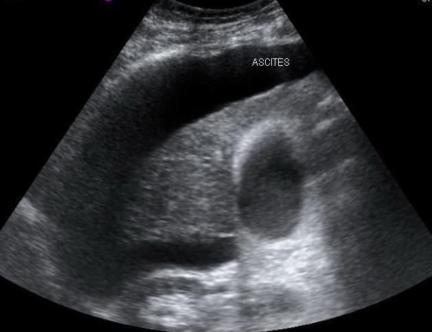
Ascites around the liver on ultrasound. The dark band labelled here is free fluid in the belly, gathered against the liver edge, a late and plain sign of the portal pressure cirrhosis builds. Fluid like this also outlines the liver’s surface, throwing any nodular bumps into relief. Image: Samir, CC BY 3.0.
A scarred liver is hard for blood to cross, and the blood backs up. The pressure that builds in the portal system is what turns cirrhosis from a quiet scar into a dangerous disease. The scan reads its marks all over the upper belly.
The spleen swells first, the surest sign of all. A spleen grown past about twelve centimetres, in a patient with a scarred liver, is the body’s gauge of the pressure behind it. The portal vein itself widens and slows; a trunk past about thirteen millimetres, carrying a sluggish flow, speaks of a system under strain. The spleen lends a number of its own where elastography is at hand: a stiff spleen tracks the portal pressure as the liver stiffness tracks the fibrosis.
The backed-up blood carves new paths. Old veins reopen, a recanalised vein running from the portal system toward the navel, and tangled collaterals appear around the stomach and the spleen, the varices that bleed. Color Doppler finds them, and finds something worse where the portal flow has turned to run backward, away from the liver it can no longer enter.
Fluid is the late, plain sign. Ascites gathers around the liver and in the flanks, a dark rim that the scan measures and tracks. A big spleen, a fat slow portal vein, collaterals, reversed flow, and free fluid together grade the portal hypertension as firmly as any single number could.
The order in which these appear is a rough grade of its own. A big spleen with a full portal vein, and nothing more, marks an early, compensated pressure. Add collaterals and reversed flow, and the system has found its escape routes. Add tense ascites and a bleed waiting in the varices, and the pressure has turned the corner into decompensation. The scan reads whether the pressure is there, and how far it has gone.
The bystanders
The liver does not suffer alone, and the scan notes its neighbours. The gallbladder wall thickens in a cirrhotic patient. The low blood protein and the portal pressure behind it do the swelling, and the gallbladder itself is healthy, a trap for the reader who takes the thick wall for cholecystitis. Fluid can climb above the diaphragm into the chest as a pleural effusion, caught on the same high view that reads the liver dome. Even the kidneys can dim and struggle when the liver fails around them. None of these grades the cirrhosis on its own. Each is a thread in the same cloth, and a reader who knows the disease looks for them as a matter of course.
A number for the hardness
The eye grades cirrhosis well at its end, less well at its start. To put a figure on the stiffness, and to catch the disease earlier, the scan reaches for elastography. The idea is simple. The harder a liver feels, the further its fibrosis has gone. The scar that drives the disease is what stiffens the tissue. The reading is taken with the patient fasted and still, several measurements pooled into one, because a single shot can mislead.
The machine sends a pulse into the liver and measures how fast the shock travels through it, faster in stiff tissue, and returns a stiffness in kilopascals. The number climbs stage by stage, from the low values of a soft liver up through the thresholds that mark advanced scarring. A reading past roughly twelve to fifteen kilopascals points toward cirrhosis, the exact threshold set by the cause.
Elastography reaches what gray-scale cannot, the early, smooth-surfaced liver that looks almost normal. Its traps are real ones: inflammation, congestion, a recent meal, or a poor patient window can all throw the number off. So the number is read beside the picture, as a companion to it. Together the look and the figure grade the liver more surely than either alone.
Different machines speak in slightly different numbers. Transient elastography and the shear-wave kind built into a regular scanner read on slightly different scales. The gap between them is small, and it matters. A reading is judged against the threshold for its own machine and the patient’s disease, never lifted bare from one setting to another. The figure is powerful, on the condition that it is read in its own language.
The liver that hides its scars
Early cirrhosis is the hardest to grade, because the liver can look nearly well. The surface has not yet broken into clear bumps. The texture is only a little coarse. The spleen sits at the top of normal. A scan read quickly here can be called clean by mistake. The cost of that miss is real, since the early liver is the one best caught, before the pressure and the cancer arrive.
Here the soft signs come into their own. A spleen creeping past its limit, a portal vein a touch full, a caudate lobe a shade large: these hints gain their force in number. In a patient with hepatitis or years of drink behind them, a clutch of them raises a flag. The early grade is built from a handful of hints, read against the patient’s story.
Reading against the last scan
Grading is not a snapshot. A run of scans, read in sequence, catches the disease in motion, often the thing that matters more than any single moment caught alone. The liver of cirrhosis is rarely still. It scars on if the cause goes untreated, and it can even soften a little once the cause is removed.
Comparison is where the soft signs harden into evidence. A spleen that has grown two centimetres since last year, a portal vein that has widened, a surface that has roughened: a change measured from one scan to the next carries a weight no single reading holds. The scan kept and measured today is the baseline that makes next year’s reading mean something. For the comparison to hold, the measurements are taken the same way each time, on the same kind of machine and the same landmarks, so a true change is told from a trick of technique.
The trajectory steers the care. A liver holding steady on treatment asks for watchful surveillance. A liver sliding deeper, its spleen swelling and its veins filling, asks for a closer eye on the pressure and the looming bleed. The grade works best as a verb. A liver is graded again and again, never only once.
The lump that must not be missed
A cirrhotic liver grows cancer. The same scarred, nodular field that makes grading hard is the soil a hepatocellular carcinoma rises from. The lumpiness that hides it is exactly why the tumor gets missed. Grading the cirrhosis and hunting the tumor are the same scan, done with both questions in mind. A cancer here often shows its hand in its blood supply. It draws a brisk arterial flow that the background nodules lack, a difference color Doppler can begin to show.
A new mass that stands out from the nodular background, a nodule that has grown since the last visit, a lump that pulls a brisk arterial flow on Doppler: each turns the scan from grading to alarm. So a graded cirrhotic liver is brought back, by the calendar, for another look, never signed off and forgotten.
The surveillance runs on a clock the grade helps set. A liver scarred enough to count as cirrhotic is rescanned every six months, often paired with a blood marker, to catch a tumor when it is still small enough to treat. The grade, in the end, is less a label than a schedule.
The nodule that turns is read for the company it keeps. A lump growing on serial scans, pulling new arteries, raising the blood marker, carries a weight no single picture gives it. The grade of the cirrhosis and the watch for the tumor run on the same clock, and the steeper the grade, the harder the watch. Catching a small cancer on a surveillance scan is why the liver was graded in the first place.
Reading it on a pocket scanner
Much of what grades a cirrhotic liver is morphology. A handheld convex scanner shows morphology exactly. The shrunken right lobe and heavy caudate, the coarse grain, the big spleen, the fat portal vein, the free fluid: a pocket device reads all of these at the bedside, and color Doppler on the better units finds the collaterals and the reversed flow. The convex probe carries the depth a fatty or a large patient demands, the same reach a cart-based scanner brings to the upper belly.
The bumpy surface asks for a high-frequency linear probe, which many handheld systems carry as a second transducer. The one thing the small machine often lacks is elastography, still largely the preserve of cart-based systems, though it is creeping into portable devices. A handheld scan can grade the established disease and its pressure with confidence; for the early, smooth liver and the precise number, it points onward.
For a clinic far from a hospital, that reach is the heart of it. A patient with hepatitis or alcohol behind them can be screened, graded, and put on a surveillance clock, on a device that travels in a bag. The scan that finds the scarred liver early is the scan that buys the patient time.
When ultrasound hands over
Ultrasound grades cirrhosis well, and it knows where its grip slips. Its grip slips on the smooth early liver, which can be read as normal when fine scarring has already begun. This is the gap elastography was made to close. Where it too falls short, a biopsy still settles the stage. A nodule is the other hand-off. The scan can flag a lump in the cirrhotic field. Saying whether it is a harmless regenerating nodule or an early cancer is the part that needs more. A suspicious nodule is sent on for a contrast study, an MRI or a CT, that reads how it takes up and washes out the dye.
The deep and the difficult belong to other tests as well. A patient too large, a belly too gassy, a liver tucked too high under the ribs can blunt the scan, and the question passes to cross-sectional imaging. Knowing when to hand over is part of grading well. The honest scan names what it could not settle and points the way on. A scan that overreaches does the real damage. Calling a stage it cannot truly see misleads more than admitting the limit and asking for a better test.
From a picture to a plan
Pull the threads together and a grade is, at bottom, an answer to one question: how far has the scarring gone, and what does that demand. The surface, the texture, the redrawn shape, the pressure downstream, and the number from elastography each add a line to that answer. No single sign carries the grade. The lines are read together, and the picture they draw is the stage the liver has reached.
A liver early in the disease asks for watchfulness and a search for the cause to halt. A liver deep in it, with a tense belly and a thicket of collaterals, asks for the management of pressure and the prevention of a bleed. Across that range, the same handful of signs, read in order and weighed together, place a patient on the map. None of it needs a particular machine, only an eye that knows the scarred liver’s marks and reads them honestly. Find the bumpy edge, weigh the spleen, follow the flow, and the scan has graded the disease as well as a picture can, on whatever device is to hand. The grade reached that way is the start of every decision that follows, from the next scan’s date to the first dose of a drug.
Common questions about grading cirrhosis on ultrasound
Can ultrasound diagnose liver cirrhosis?
It can, especially once the disease is established. A nodular liver surface, a coarse texture, a shrunken right lobe with an enlarged caudate, and the signs of portal hypertension together make the diagnosis with confidence. Early cirrhosis, with a near-normal liver, is harder, and elastography or a biopsy may be needed to settle it.
Which ultrasound sign of cirrhosis is the firmest?
A frankly nodular liver surface is among the more specific. It is seen best with a high-frequency linear probe over the front of the liver, and it shows clearest when a little ascites outlines the bumps. The caudate-to-right-lobe ratio and the signs of portal hypertension support it.
How does elastography grade liver fibrosis?
It measures the liver’s stiffness, since a scarred liver is harder than a healthy one. A pulse is sent in and its speed through the tissue is read as a stiffness in kilopascals. The figure climbs stage by stage, and a value past roughly twelve to fifteen kilopascals points toward cirrhosis, the exact threshold set by the cause.
What ultrasound signs show portal hypertension?
An enlarged spleen, a dilated and slow portal vein, collateral veins around the stomach and toward the navel, free fluid in the belly, and a portal flow that has reversed to run away from the liver. The spleen size and the reversed flow are among the firmest of these.
Why is a cirrhotic liver scanned every six months?
Because cirrhosis is the soil that liver cancer grows in. A six-monthly scan, often with a blood marker, aims to catch a hepatocellular carcinoma when it is small and treatable. The nodular liver that makes grading hard also hides tumors, so the repeat scan is read with the cancer in mind.
Can a handheld ultrasound grade cirrhosis?
For the established disease, yes. A handheld convex scanner shows the shrunken lobe, the coarse texture, the big spleen, the full portal vein, and the free fluid. Doppler finds the collaterals and reversed flow. The early smooth liver and the exact stiffness number usually need elastography. The typical handheld unit still lacks it.
About the Author
Julien Mercier
Senior R&D Engineer · Medical Ultrasound Transducer Development
Senior R&D Engineer with an M.S. in Applied Physics and over 15 years of experience in medical ultrasound transducer development, specializing in the design verification and performance testing of high-frequency imaging transducers. Currently leading the development and verification of the company’s next-generation high-frequency linear-array transducer, responsible for imaging performance evaluation and reliability analysis in preclinical testing. Brings extensive hands-on experience in piezoelectric element tuning, beamforming parameter optimization, and system-level performance testing.