Shoulder Joint Ultrasound Scanning Handheld Linear Probe
Scanning the shoulder with ultrasound means working through a fixed run of arm positions, one rotator-cuff tendon at a time. No single position shows all four tendons, because each one tucks behind bone until the arm is moved to bring it into view. The order of positions stays the same on every patient. A position dropped is a tendon dropped with it, and a tear in the part left out gets signed off as a normal shoulder.
What the shoulder scan covers
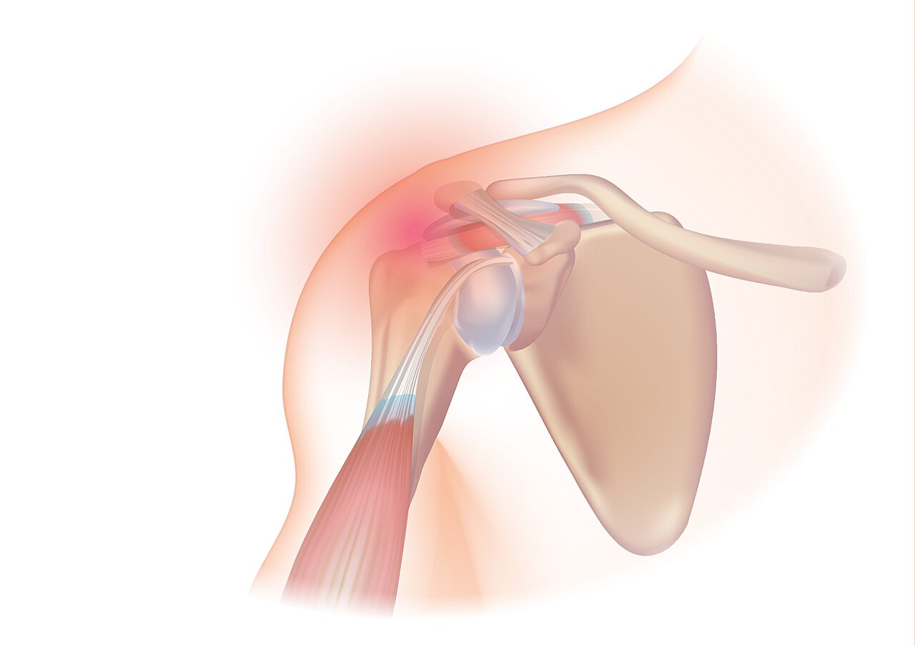
The shoulder seen from the front. The rotator cuff wraps over the head of the upper arm bone and passes under the bony arch above, with the collarbone and the AC joint across the top. The red glow marks the subacromial region, where the cuff is pinched and impingement pain begins. Illustration: Injurymap, CC BY 4.0.
The rotator cuff is four tendons. The supraspinatus lies on top, the infraspinatus and the small teres minor behind, the subscapularis in front. Each runs from the shoulder blade to the head of the upper arm bone. Together they hold the joint in place and turn it.
Above the cuff sits the subacromial bursa, a thin sac that lets the tendons glide under the bony arch of the shoulder. In front, the long head of the biceps tendon runs down a groove in the bone. The joint between the collarbone and the shoulder blade, the AC joint, sits over the top. These are the structures a full scan works through.
Each structure has a position that brings it into view. The cuff tendons converge onto the head of the bone from the front, the top and the back, each meeting it at its own angle. That spread is why no single placement of the probe catches them all. The arm is moved to a fresh position to bring each tendon forward in turn.
The order matters because the cuff is a continuous sheet, one tendon blending into the next. A tear often starts in one tendon and runs into the neighbor. Reading the cuff in sequence shows where one tendon ends and where a tear has crossed into the next. A scan that jumps straight to the painful spot can miss the edge of a tear in the tendon beside it.
The biceps tendon, a place to start
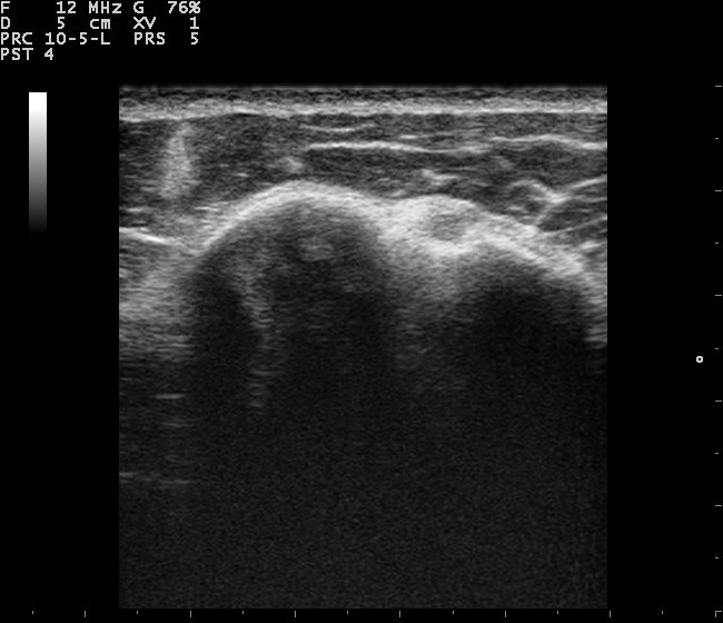
The biceps tendon in short axis, a cross-view at the front of the upper arm. The tendon sits as a rounded structure in its groove, the notch between the two bony bumps of the humeral head. The text at the top left is the scanner’s settings, not part of the anatomy. Image: Nevit Dilmen, CC BY-SA 3.0.
The biceps tendon is a good place to start, easy to find and a fixed landmark for the rest. With the arm resting on the thigh, palm up, the probe is laid across the front of the upper arm. The tendon shows in short axis as a bright oval, sitting in its groove between two bony lips.
Turned ninety degrees, the probe shows the same tendon in long axis, a bright fibrillar band running down the groove. Both planes are scanned. A split tendon, or one slipped out of its groove, shows in one plane more plainly than the other. A little fluid around the tendon in its sheath can point to trouble higher in the joint.
The groove is a landmark. The subscapularis tendon lies to the inside of it, the supraspinatus directly above. Finding the biceps first orients the operator for the tendons that follow.
A biceps tendon thickened, or surrounded by fluid, signals wear, or a problem in the joint it runs from. An empty groove, with the tendon gone from it, means the tendon has slipped out or torn through. The short-axis view catches an empty groove at once.
The state of the biceps often hints at the cuff above it.
The subscapularis in front
The subscapularis is the cuff tendon at the front. It comes into view when the arm is rotated outward. With the elbow held at the side and the forearm turned out, the tendon swings under the probe laid across the front of the shoulder. The rotation draws it from behind the bone, where it would otherwise hide.
In this transverse view the tendon shows its fibrillar fingers running to the lesser tubercle, the bony bump it attaches to. The probe slides up and down the tendon, then turns along it, to check its length in both planes. The outward rotation is the step that matters. Leave it out, and a tear stays hidden behind the bone.
The subscapularis tears less often than the tendon on top. It still carries part of the cuff’s work. A scan that skips it for the supraspinatus alone leaves a quarter of the cuff unchecked.
The lesser tubercle turns to the front as the arm rotates out. Watching the bone turn under the probe confirms the right structure is in view. A subscapularis tear shows as a gap at this attachment, sometimes with the biceps tendon slipped from its nearby groove. The two often occur together.
The supraspinatus, the central tendon
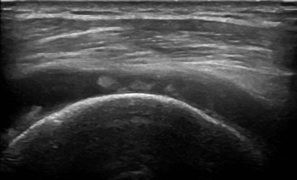
A rotator cuff scanned in long axis over the curved head of the upper arm bone, from a case recorded as a cuff tear. A tear shows where the even bright band of the tendon gives way to a darker gap above the bone. Image: Mme Mim, CC BY-SA 3.0.
The supraspinatus is the tendon a shoulder scan centers on, and the one of the four likeliest to tear. It runs through a tight gap under the bony arch, where it is pinched and where it takes much of the strain each time the arm lifts. Bringing it into clear view takes a particular position. The patient brings the hand behind the back to rest on the hip, in the back-pocket position, with the elbow drawn backward. This is the modified Crass position. It swings the supraspinatus forward from under the bony arch, where the tendon otherwise hides. The move lays the tendon flat under the probe. In long axis the supraspinatus shows as a curved fibrillar band running to its footprint on the bone. That curve is what makes anisotropy such a trap here. As the tendon bends, the beam falls square on only part of it at a time. The probe is rocked along the curve to keep each stretch bright, a habit the beginner’s page covers in full. Turned ninety degrees, in short axis, the tendon is swept from front to back, since a tear can sit in one part and spare the rest. A tear in the supraspinatus is the finding the scan is built to catch. It shows as a defect in the curved band, a spot where the fibrillar pattern stops and fluid or an empty gap takes its place. A tear may thin one surface of the tendon, break clean through the full thickness, or open a gap wide enough for the muscle to draw the torn end back toward the shoulder blade. The footprint, where the tendon meets the bone, is a common site for a tear, so the insertion is watched closely as the probe sweeps across it. Comparing the tendon’s thickness and brightness against the other shoulder helps, since a worn supraspinatus can be subtle on its own. Through all of this the probe stays square to the curving tendon. The anisotropy that darkens a tilted stretch can mimic a tear that is not there. It can hide a real one in the shadow it casts. Calcification gathers in the supraspinatus more than in any other cuff tendon. Reading the tendon takes both planes, a steady square beam, and a patient placed to bring the tendon out. Scanning the full supraspinatus, end to end and in both planes, is the center of the shoulder examination.
The infraspinatus and teres minor behind
Behind the shoulder lie the infraspinatus and, below it, the small teres minor. They come into view with the arm brought across the body, the hand reaching to the opposite shoulder. That movement turns the back of the cuff forward. The probe then sits just below the spine of the shoulder blade, in a long view of the posterior cuff.
Tears at the back of the cuff are the less common kind. A scan that stops at the top still misses them. The infraspinatus is the one to check of the two, since it shares in many of the same problems and lies within easy reach from behind. The back of the cuff belongs to a full examination.
The same posterior view reaches the back of the main shoulder joint, where the arm bone meets its socket. A rim of cartilage, the labrum, sits at the edge, hard to see in full from outside. Fluid pooling in this posterior recess is one of the clearer signs of a joint that is inflamed or injured, beyond the trace a normal shoulder can hold.
The AC joint on top
The AC joint, where the collarbone meets the shoulder blade, sits at the top of the scan. The probe is laid across the top of the shoulder, over the gap between the end of the collarbone and the bony point of the shoulder. A normal joint reads as a thin gap with a small disc of cartilage inside. It is quick to reach and easy to pass over in a hurry.
Wear in this joint is common and often missed as a source of pain. It shows as a rough, built-up bone edge, the joint capsule swollen into a bulge that lifts the skin above it. A scan that reads the cuff in full includes the AC joint at the top. Pain felt over the top of the shoulder can come from this joint as much as from the cuff below it.
What the shoulder scan looks for
A tear is the main finding the scan looks for. It reads as a gap in the fibrillar band, a spot where fluid or nothing has taken the place of tendon. The scan grades it by the depth of the gap and by whether the torn ends have drawn apart.
Calcification shows as a bright spot or streak inside a tendon, usually the supraspinatus, with a shadow trailing behind a dense deposit. The subacromial bursa, normally a thin line, reads as thickened or fluid-filled when it is inflamed, the rubbing called impingement. Tendinopathy, short of a tear, shows as a tendon swollen and darker than normal, its fibrillar pattern dulled.
Color Doppler adds one more sign. A tendon or a bursa lit with color where it should be quiet points to the raised blood flow of active inflammation. Read together, these signs separate a cuff that is intact from one that is torn, inflamed or worn.
The size of a tear guides what comes next. A small partial tear is often left to heal without surgery. Repair is the usual path for a large, full-thickness tear with the ends drawn back. The scan measures the gap and notes whether the torn ends can still be seen, since a tendon retracted far back is harder to fix.
The scan in order
The views run in a set order so that none is forgotten. A common sequence starts at the AC joint on top, moves to the biceps tendon in front, then to the subscapularis with the arm rotated out, then to the supraspinatus in the modified Crass position, and finishes with the infraspinatus and teres minor behind. The order can vary. A fixed one, followed every time, guards against a missed tendon.
Dynamic views are added where a problem is suspected. The arm is moved through lifting and rotation. The probe follows the supraspinatus and the bursa under the bony arch. A still scan can miss an impingement that shows only when the arm moves. The same movement can bring out a biceps tendon that slips from its groove, or a bursa that bunches as the arm rises.
The sore spot is only part of the scan. Working through the rest of the cuff each time catches the damage the painful spot hides, since pain in one place can come from a tear in another.
Each view is held long enough to read. A tendon caught for only a moment can hide a tear that a steady look would show. An image saved at each step builds a record, a baseline for the next scan to compare against.
Shoulder ultrasound on a handheld unit
The shoulder suits a handheld scan. The cuff tendons are shallow, within easy reach of a linear probe. The patient can be moved through the scanning positions in a chair, with no need for a fixed room.
The handheld form brings the scan to the clinic or the sports field, where a painful shoulder can be checked on the spot and compared against the other side. The dynamic views, where the arm moves through its range, work as well at the bedside as anywhere. The same routine and the same positions apply, whatever the size of the machine.
What the small unit asks is a careful, ordered technique: each tendon in its position, both planes, and the dynamic views where they help. Run in full, that technique gives a sound scan on a handheld unit as on any in the department.
Common questions about shoulder ultrasound
What does a shoulder ultrasound show?
It shows the rotator cuff, the four tendons around the joint, along with the long head of the biceps tendon, the subacromial bursa and the AC joint. Each is brought into view with the arm in its own position. The scan reads them for tears, calcification and inflammation.
How is the supraspinatus scanned?
With the hand brought behind the back to rest on the hip, the back-pocket position, and the elbow pointing back, the modified Crass position. This swings the supraspinatus out from under the bony arch. It is then scanned in long and short axis, with the probe rocked along its curve to keep the tendon bright.
Why does the scan center on the supraspinatus?
It is the cuff tendon likeliest to tear, and it runs through a tight gap under the bony arch where it is pinched and bears much of the strain as the arm lifts. The scan reads it end to end, in both planes, and against the other shoulder.
What problems can shoulder ultrasound find?
Tears of the cuff tendons, both partial and full-thickness, along with calcification in a tendon, inflammation and fluid in the subacromial bursa, tendinopathy and impingement seen on dynamic views. Color Doppler adds the raised blood flow of active inflammation.
Why scan the shoulder in a set order?
The cuff tendons curl around the head of the bone, so each needs its own arm position and no single view shows them all. A fixed order, run the same way each time, stops a sore spot from pulling the scan away before every tendon is seen.
Can a handheld ultrasound scan the shoulder?
Yes. The cuff tendons are shallow and within reach of a linear probe. The patient can be moved through the positions in a chair. A handheld unit also does the dynamic views at the bedside or on the field, with the other shoulder a moment away for comparison.
About the Author
Julien Mercier
Senior R&D Engineer · Medical Ultrasound Transducer Development
Senior R&D Engineer with an M.S. in Applied Physics and over 15 years of experience in medical ultrasound transducer development, specializing in the design verification and performance testing of high-frequency imaging transducers. Currently leading the development and verification of the company’s next-generation high-frequency linear-array transducer, responsible for imaging performance evaluation and reliability analysis in preclinical testing. Brings extensive hands-on experience in piezoelectric element tuning, beamforming parameter optimization, and system-level performance testing.