A radial line is not there to give fluids. It reads blood pressure beat by beat and draws blood for gas analysis, the two jobs a vein cannot do, through a catheter in the small artery at the wrist. The artery is easy to feel in many people; when the pulse is faint or the artery rolls away, ultrasound shows it and guides the cannula in.
What an arterial line is for
An arterial line does two things a peripheral drip cannot. It reads the blood pressure straight from inside the artery, beat by beat, shown as a live waveform. A cuff gives only a single number every few minutes. In a patient whose pressure is swinging, that continuous reading is the point.
The waveform carries more than a number. Its shape tells the trained eye about the heart’s output and the stiffness of the arteries. The peak and trough of each beat give the systolic and diastolic pressure, with the mean read off as well. A cuff gives none of this between its readings.
The second use is blood sampling. An arterial line lets blood be drawn for gas analysis without a fresh needle each time, which matters in a patient who needs gases checked often, on a ventilator or in shock. The line is drawn from again and again, with no new stick.
None of this is what a vein line does. A vein line carries fluids and drugs into the body. The artery line works the other way, reading pressure and drawing blood out. The two go in separate vessels for separate reasons. Confusing them is dangerous, since a drug meant for a vein can harm an artery.
Reading the arterial trace
The line is only as good as the trace it produces. A clean arterial waveform rises sharply with each beat to a clear peak. On the way down it shows a small notch where the aortic valve closes. The shape reports on the circulation, a sharp tall trace in a strong heart, a low slurred one in a failing heart.
The trace can mislead when the tubing is wrong. Air bubbles or a soft line damp the signal, flattening the peak and reading the pressure too low. A stiff or clotted line does the reverse, throwing sharp spikes that read too high. A quick flush test, watching how the trace settles, tells whether the reading can be trusted.
Why the radial artery is the chosen site
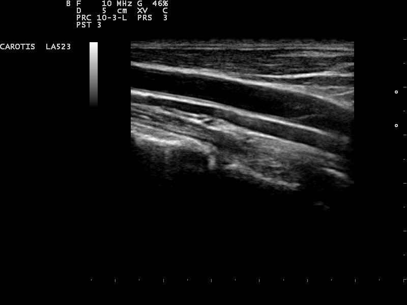
An artery on ultrasound in long axis, here the carotid in the neck. The radial at the wrist runs the same way, close beneath the skin.
The radial artery is the usual choice for an arterial line. It runs close under the skin at the wrist, easy to reach with a short cannula. It lies away from the deeper structures that a more central artery would put at risk.
Safety is the larger reason. The hand is fed by two arteries, the radial and the ulnar, joined in arches across the palm. If the radial is harmed or blocked by the line, the ulnar can supply the hand on its own. This double supply is why the wrist is a safer site than an artery that feeds its tissue alone.
The numbers bear out the safety. Radial artery catheterization carries a rate of permanent ischemic harm around 0.09 percent, roughly one in a thousand. That is low enough to make the radial the first choice at the great majority of centers. The rare harm comes when the backup supply was poor to begin with.
Other arteries can take a line when the radial cannot. The femoral artery in the groin is larger and easier in a collapsed patient. It sits deeper and closer to harm. The brachial at the elbow and the dorsalis pedis on the foot are used at times. The radial stays first for its mix of access and safety, with the others kept for when it fails.
Why ultrasound beats the finger
A radial line often goes in by feel, the finger on the pulse guiding the needle. This works when the pulse is strong. It fails when the pulse is faint, in a patient who is cold or in shock, whose pulse the finger cannot find. These are often the patients in greatest need of the line. Ultrasound shows the artery the finger cannot. The numbers from trials are consistent. One randomized trial in elderly patients reported a first-attempt success of 84 percent with ultrasound against 56 percent by palpation. A trial in a pre-anesthesia room reported 97 percent against 73 percent. A large randomized trial of around 750 patients found ultrasound lifted the first-attempt success by roughly 14 points over palpation. The exact figures vary with the patient and the study. Every trial points the same way, ultrasound ahead of the finger. The gain is largest where the pulse is weakest. Where the finger has little to work with, in a child or a shocked adult, the screen shows the artery plainly. A trial in children found the time to a working line cut from over 10 minutes to around 3, with far fewer complications along the way. The faster line is more than convenience. A patient in shock needs the pressure reading without delay. Minutes saved at the wrist are minutes the rest of the care is not kept waiting. Seeing the artery does more than find it. The depth and the angle are read before the needle moves, the spot chosen where the artery runs straight and wide, away from a branch. The needle is watched into the vessel, no longer pushed toward a pulse that may have shifted. One refinement helps with so small a target. The tip is kept in view and walked forward in small steps, the probe sliding ahead of it, so the tip is always the part being watched as it nears the artery. This dynamic tracking suits a vessel that a single thrust would push through. The artery also moves under the needle, rolling aside or diving deeper. The finger cannot track that. The probe can follow it. For the hard stick, ultrasound often lands the line in one pass where the finger needs many, so the patient feels fewer needles and the line is working sooner. Each failed pass can bruise the artery or set it into spasm. Fewer passes mean a cleaner vessel for the line that finally goes in. For a difficult wrist, the screen is what makes the line possible at all.
The Allen test and the backup supply
Before a radial line goes in, the hand’s backup supply is often checked. The aim is to be sure the ulnar artery can feed the hand if the radial is later blocked. The classic check is the modified Allen test. Both wrist arteries are pressed, the hand is squeezed pale, and the ulnar alone is released to see whether color floods back.
The Allen test is widely done. Whether it works is another matter. The test is meant to predict who will lose blood supply if the radial closes, and the evidence that it predicts this is weak. A hand that fails the test may come to no harm. A pass is no guarantee of safety either. Many operators run it from habit and caution more than from proof.
Ultrasound offers a firmer check. The probe can watch the ulnar flow directly. A dynamic version measures the ulnar speed at the wrist before and after the radial is pressed, a rise in flow showing the ulnar can take over. This reads the backup supply by what it does. The color of a squeezed hand is a cruder sign.
A pulse oximeter on the thumb gives another read, the Barbeau test, where the trace is watched as the radial is pressed. A trace that holds shows the ulnar is carrying the hand. This needs no help from the patient, useful in someone sedated or unwell.
The artery is not a vein
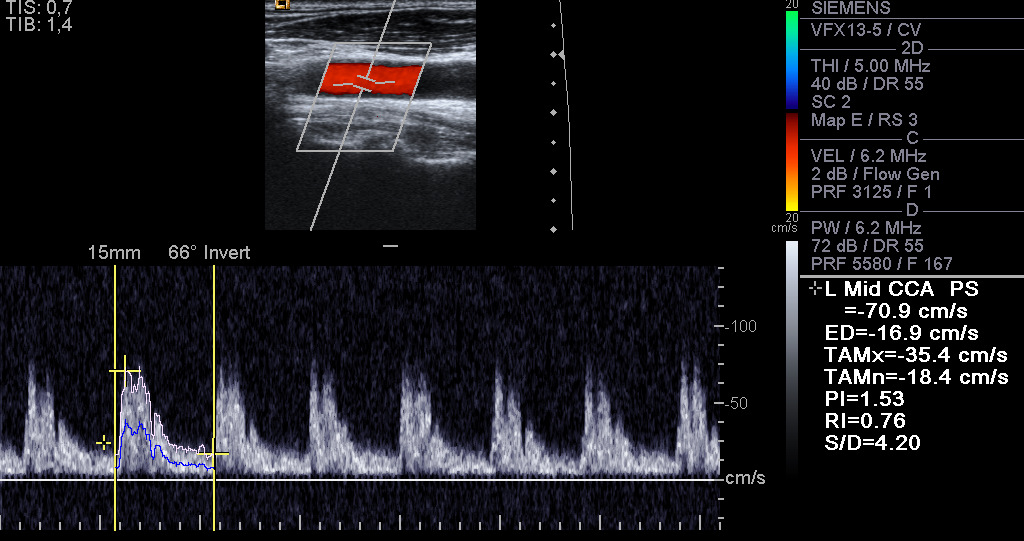
An artery with colour and spectral Doppler, here the carotid. The fast, pulsing flow marks an artery, the sign that tells the radial from a vein at the wrist.
On the screen the artery has its own look. The difference from a vein guides the stick. The artery is round and stays round under the probe, where a vein would squash flat. It pulses with the heartbeat. It sits thick-walled and bright-rimmed. Color Doppler fills it with the fast, pulsing flow of an artery.
The look is the easy part.
The radial artery brings its own challenges. It is small, only a few millimeters across, a narrow target even on the screen. It is prone to spasm, clamping down when the needle touches it, which shrinks the target further. A gentle, steady approach matters more here than in a wide vein.
Spasm is handled in two ways. Warming the arm and numbing the skin both lower the chance of it, as does an unhurried needle. The artery is given time to relax if it clamps down. Forcing a spasmed artery only makes it worse.
The same care that finds the artery avoids its neighbors. The radial nerve runs near the artery low on the forearm. A vein may lie beside it. The probe sorts them out, the artery known by its pulse, so the needle goes to the right vessel.
Guiding the cannula and avoiding occlusion
The cannula goes in under live view, the artery held on the screen and the needle brought to it. The way the needle lines up with the probe, across the beam or along it, is its own technique with its own pages. What counts here is that the artery is watched, the pulse no longer the only guide.
A flash of bright red blood, pulsing back, marks the artery entered. The cannula is threaded off the needle into the vessel. With the artery small and its walls springy, the operator advances gently, since a rough push can tear the back wall or send the artery into spasm.
The main late problem is the artery closing off. A catheter that fills too much of the narrow radial slows the flow past it and invites a clot. Ultrasound has measured this radial occlusion at higher rates than once thought, more so with a larger catheter. Reading the artery’s size on the screen lets the operator match the catheter to it, leaving flow around the line and lowering the risk.
The catheter for a radial is small, sized to the artery and smaller than a venous cannula. When the line comes out, firm pressure is held on the wrist for several minutes, longer than a vein would need, since an artery bleeds under pressure. The same probe can check afterward that the flow has returned and no clot has blocked the vessel.
After the line goes in
An arterial line needs watching while it stays in. The hand is checked for color and warmth, the signs that blood is still reaching it. A hand that turns pale or cold warns that the radial has closed and the ulnar is not keeping up. That is a reason to pull the line.
The line is flushed slowly to keep it from clotting. It is labelled clearly as arterial, since a drug pushed into it by mistake can harm the hand. When it is no longer needed it comes out. Pressure is held until the bleeding stops.
The probe has a last role at removal. Once the line is out and the pressure released, a quick scan checks that the radial has reopened and blood runs through it again. A radial found blocked is followed up, since the hand is living on the ulnar alone.
A blocked radial often opens again on its own over the days that follow. One that stays shut is watched by scan until the hand is known to be safe.
Placing an arterial line on a handheld unit
A handheld unit places the arterial line wherever the patient is, from theatre to intensive care. The probe finds the artery, checks the backup flow, then guides the cannula, all at the wrist without extra kit. A faint pulse no longer blocks the line.
Common questions about radial arterial lines
What is a radial arterial line used for?
It reads the blood pressure beat by beat as a live waveform and lets blood be drawn for gas analysis without a fresh needle each time. It carries no fluids or drugs, which is the job of a vein line. It is used in surgery and intensive care.
Why is the radial artery the usual site?
It runs close under the skin at the wrist, and the hand has a second artery, the ulnar, that can supply it if the radial is blocked. This backup makes the wrist safe, with permanent ischemic harm around 0.09 percent.
Does ultrasound improve radial artery cannulation?
Yes, especially when the pulse is faint. One trial in elderly patients reported first-attempt success of 84 percent with ultrasound against 56 percent by palpation. A trial in children cut the time to a working line from over 10 minutes to around 3.
Is the Allen test needed before a radial line?
The modified Allen test checks the ulnar backup supply and is widely done. The evidence that it predicts ischemic harm is weak. Ultrasound can check the ulnar flow directly, a firmer reading than the color of a squeezed hand.
What sets the artery apart from a vein on ultrasound?
The artery is round and stays round under probe pressure, where a vein squashes flat. It pulses with the heartbeat and shows fast, pulsing flow on color Doppler. The radial artery is small and prone to spasm.
Can a handheld ultrasound place an arterial line?
Yes. A handheld probe shows the artery, checks the ulnar backup flow, and guides the cannula into the wrist in real time. The line is placed at the bedside, useful when the pulse is too faint to feel.
About the Author
Julien Mercier
Senior R&D Engineer · Medical Ultrasound Transducer Development
Senior R&D Engineer with an M.S. in Applied Physics and over 15 years of experience in medical ultrasound transducer development, specializing in the design verification and performance testing of high-frequency imaging transducers. Currently leading the development and verification of the company’s next-generation high-frequency linear-array transducer, responsible for imaging performance evaluation and reliability analysis in preclinical testing. Brings extensive hands-on experience in piezoelectric element tuning, beamforming parameter optimization, and system-level performance testing.