Axillary Lymph Node Ultrasound Assessment Handheld Linear Probe
The axilla is the first place breast cancer spreads. Cancer cells leaving a breast tumor travel to the lymph nodes under the arm before they reach anywhere else, which is why checking those nodes is part of staging the disease. A normal node and a node that may hold tumor look different on ultrasound, and telling them apart shifts both the stage and the treatment that follows.
The normal axillary lymph node
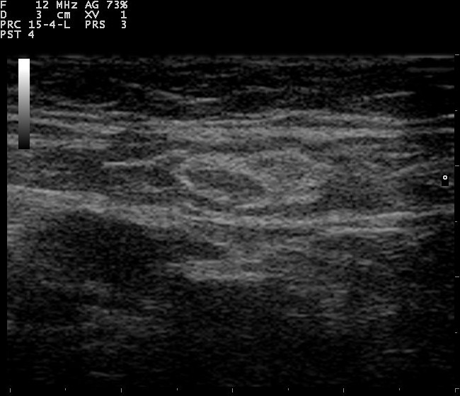
A lymph node on ultrasound showing a thin cortex around a bright fatty hilum, the normal build an axillary node shares.
A normal lymph node has a shape worth knowing, since the shape is what changes with disease. On ultrasound it looks like a small kidney bean. The outer rim is the cortex, a thin dark layer of lymphoid tissue. The center is the hilum, a bright zone of fat where the vessels enter and leave. The cortex of a normal node stays thin, under 3 millimeters. It runs even all the way around.
The bright fatty hilum is the reassuring sign. A node with a clear fat center and a thin even rim is almost always benign. Some normal nodes hold so much fat that they fade into the fat of the armpit around them, hard to pick out, which is itself a sign of health.
Size alone says little. A reactive node fighting an infection can grow large and still keep its normal shape, its fatty hilum and its thin cortex. The reader looks at the build of a node before its size. A big node with a normal structure is calmer than a small one with a thick cortex.
A node can change for harmless reasons. An infection in the arm or hand can swell the nodes that drain it. So can a recent vaccination or a healing cut. They settle back once the cause passes. History counts as much as the picture here. A node swollen by a recent infection is read against that cause. One found beside a breast tumor with no other reason draws more concern.
What metastasis does to a node
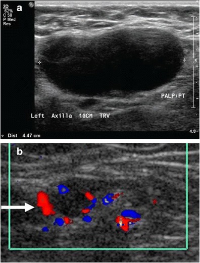
Metastasis changes the node from the cortex inward. Tumor cells arrive through the lymph vessel that feeds the node and lodge in the cortex first, since the cortex is where the node filters what the lymph carries. The cortex thickens where they settle, at first in one spot, a focal bulge on one side with the rest still thin. This eccentric thickening is an early sign, one a reader learns to catch before the rest of the node looks wrong. As more cells arrive the cortex swells around more of the rim, until it has lost its thin even line all the way around. As the tumor grows it pushes on the fatty hilum. The bright fat center is squeezed to one side, then lost altogether as the cortex fills the node. A node that has lost its fatty hilum is the strongest warning sign on the scan. The absent hilum carries a positive predictive value of 86 to 90 percent for metastasis. Shape shifts along with the inside. A normal node is longer than it is wide, an oval. A node filling with tumor rounds out, growing as wide as it is long. Blood flow can add to the picture. A normal node draws its blood through the hilum, so color Doppler shows flow entering at the fat center. A node taken over by tumor grows new vessels at its edge, so the flow moves to the cortex and the rim, scattered and out of the orderly hilar pattern. Tiny bright flecks of calcium inside a node, less common than the other signs, point the same way, since some breast cancers carry calcification into the nodes they reach. No single sign is proof on its own. A node can hold a small deposit and still look nearly normal, which sets the floor on what the scan can promise. The biggest nodes are not always the worst. A reactive node can be large with a clean structure. A small node packed with tumor can carry every bad sign. The reading is comparative as much as absolute, since a node that stands out from its neighbors in cortex or shape draws the eye before any measurement. What looks like one large node can also be several matted together, a sign of heavier disease. The reader weighs all of these together. A node that fails on several counts is the one sent for a needle.
The cortical thickness measurement
A suspected malignant axillary lymph node. The grayscale image shows a rounded node that has lost its fatty hilum. Color Doppler shows disordered flow at the rim.
Of all the signs, cortical thickness is the one that can be measured. A reader can put calipers on the cortex and read a number, where shape and hilum are judged by eye. The number gives the assessment something firm to stand on.
The cortex is where the change shows first.
The threshold in common use is 3 millimeters. A cortex thicker than 3 millimeters is the single sharpest sign of a node holding tumor. A node with a cortex over that line carries around four times the risk of metastasis compared with a node under it. The numbers behind the threshold are stark. Metastatic nodes in one study ran a median cortical thickness of 7.5 millimeters, against 2.1 millimeters for normal nodes. A cortex of 3 millimeters or more turned up in 83 percent of the cancerous nodes and in only 17 percent of the clear ones.
The measurement is taken at the thickest part of the cortex, since tumor often thickens it on one side first. Reading both armpits and comparing helps, because a woman’s two sides usually match, so a thicker cortex on the cancer side stands out against her own normal node across the body. High-frequency harmonic imaging, in the 11 to 15 megahertz range, sharpens the cortex enough to measure it well. An average reading around the node would hide the single bulge a deposit makes, which is why the thickest point is what matters. A focal thickening counts even when the rest of the cortex stays thin.
Where the axilla is scanned
The axilla is scanned in a set order so no group of nodes is skipped. The nodes sit in levels, named by where they lie against a chest muscle called pectoralis minor. Level one is below and to the outside of the muscle, level two behind it, level three above it toward the collarbone.
The cancer reaching the axilla lands in level one first as a rule, so that is where the scan starts and looks hardest. It then works up through levels two and three. A node out of order, high in the axilla with the lower levels clear, is unusual and gets noted.
The probe sweeps the hollow of the armpit with the arm raised, which opens the space and brings the nodes closer to the skin. The high-frequency linear probe suits the job, since the nodes sit shallow. The operator angles up behind the muscle to reach the higher levels, where the nodes are harder to bring into view.
A few nodes sit off the main levels. Rotter’s nodes lie between the two chest muscles. The internal mammary nodes run beside the breastbone, reached by angling the probe between the ribs. These are checked when the main levels raise concern or when the tumor sits where it would drain to them.
The limits of the scan
Ultrasound does not catch every involved node. Its sensitivity for nodal metastasis runs around 62 percent, so it misses roughly a third of the nodes that turn out to hold tumor. The misses are usually small deposits, a few cells or a tiny focus that has not thickened the cortex or changed the shape.
These small deposits have names set by size. A micrometastasis runs from above 0.2 millimeters up to 2 millimeters. Isolated tumor cells are smaller still, at 0.2 millimeters or less, sometimes only a few cells. Neither swells the cortex enough to show on a scan. Both are found only when the node is taken out and read under the microscope.
Their meaning is still debated. The smallest deposits may carry less weight than a fully involved node. The size cutoffs were drawn partly to help pathologists agree with one another.
What ultrasound does well is the other side, ruling cancer in. Its specificity sits near 93 percent, so a node that looks clearly abnormal on the scan is likely to hold tumor, with few false alarms. A negative predictive value near 82 percent means a normal-looking axilla is reassuring without being a guarantee.
This is why the sentinel lymph node biopsy stays the standard for staging. The sentinel node is the first node the breast drains to, found by a tracer and removed for the pathologist to read. Ultrasound does not replace it. Ultrasound finds the nodes worth sampling before surgery. A positive needle can move a patient straight to fuller treatment, skipping the sentinel step.
What the node count adds up to
The number of nodes with tumor sets the nodal stage. One to three involved nodes counts as stage N1. The stage rises to N2 at four to nine nodes. Ten or more makes it N3. The count feeds the overall stage that steers treatment.
This is why the scan tallies the nodes on top of judging each one. A lone suspicious node points lower on that scale than a cluster of them. The ultrasound gives an early read on how far the disease has reached before any node is removed. A scan that points to heavy nodal disease can move the plan toward chemotherapy first.
From a suspicious node to a needle
A node that looks abnormal goes to a needle before any surgery. The same probe that found it guides the sample. A fine needle can draw cells from the thickened cortex, or a core needle can take a small piece, the same way a breast mass is sampled. The cortex is the target, since that is where the tumor sits. The fatty hilum holds none.
A positive result settles the question before the operation. It tells the surgeon the axilla is involved, which can change the surgery from a sentinel sampling to a fuller clearance, or move chemotherapy ahead of surgery. The needle turns a suspicious picture into a fact to plan around.
The needle carries a small risk of its own. Spreading tumor along the track is a theoretical worry, held low by taking the shortest safe path and by the shallow depth of the node.
Reading the axilla after chemotherapy
Chemotherapy given before surgery can clear the nodes of tumor. A node that held cancer at diagnosis can look normal again after treatment, its cortex thinned and its hilum back. Checking the axilla after chemotherapy shows how well the treatment worked.
This raises a problem of memory. A node that has responded looks like a node that was never involved, so the spot needs marking before treatment starts. A tiny clip placed in the suspicious node at the first biopsy lets the surgeon find that exact node later, after the cancer in it is gone.
Removing the marked node along with the sentinel nodes checks the response where it matters. A clip in the node, placed under ultrasound, makes that targeted check possible. The scan that staged the axilla at the start guides the marking that reads the result at the end.
The response read this way guides what comes next. A node cleared of tumor can let the surgery stay smaller. Nodes still involved point to a fuller clearance. The marked node, read after treatment, is the one that settles how much surgery the axilla still needs.
Assessing nodes on a handheld unit
A handheld unit scans the axilla in the same sitting as the breast. The operator sweeps the levels with the high-frequency probe, measures any thick cortex, then checks each node for its fatty hilum. A suspicious node can be sampled on the spot. The portable form brings nodal staging to the clinic.
Common questions about axillary lymph node ultrasound
Why is the axilla checked in breast cancer?
The axilla is the first place breast cancer tends to spread. Whether the lymph nodes under the arm hold tumor changes the stage, the surgery, and the rest of the treatment, so the nodes are checked as part of working up a breast cancer.
What does a normal axillary lymph node look like on ultrasound?
It looks like a small kidney bean, with a thin dark cortex under 3 millimeters around a bright center of fat called the hilum. A clear fatty hilum and a thin even rim mark a node as almost always benign.
What ultrasound features suggest a lymph node holds cancer?
A cortex thicker than 3 millimeters is the main warning sign. A lost fatty hilum, a round shape and blood flow out at the rim add to it. An absent hilum alone carries an 86 to 90 percent predictive value for metastasis.
How accurate is axillary ultrasound for finding cancer in the nodes?
Its sensitivity is around 62 percent, so it misses some small deposits. Its specificity is around 93 percent, so an abnormal node is likely to hold tumor. A cortex over 3 millimeters carries about four times the risk of metastasis.
Does ultrasound replace sentinel lymph node biopsy?
No. The sentinel lymph node biopsy stays the reference standard for staging the axilla. Ultrasound finds the nodes worth sampling before surgery, and a positive needle can move a patient toward fuller treatment ahead of the sentinel step.
Can a handheld ultrasound assess axillary lymph nodes?
Yes. A handheld high-frequency probe sweeps the axillary levels, measures the cortex of each node, and checks for a fatty hilum, all in the same sitting as the breast scan. A suspicious node can be sampled on the spot.
About the Author
Julien Mercier
Senior R&D Engineer · Medical Ultrasound Transducer Development
Senior R&D Engineer with an M.S. in Applied Physics and over 15 years of experience in medical ultrasound transducer development, specializing in the design verification and performance testing of high-frequency imaging transducers. Currently leading the development and verification of the company’s next-generation high-frequency linear-array transducer, responsible for imaging performance evaluation and reliability analysis in preclinical testing. Brings extensive hands-on experience in piezoelectric element tuning, beamforming parameter optimization, and system-level performance testing.