Breast Nodule Benign Versus Malignant Ultrasound Differentiation Handheld
Telling a benign breast nodule from a cancer is the main job of breast ultrasound. The large majority of breast nodules are benign, a fibroadenoma or a cyst that needs no treatment, while a few are cancers where early detection changes everything. Ultrasound reads the nodule and sorts it toward one side or the other by the way it looks and the way it behaves, feature by feature.
How ultrasound separates benign from malignant
Ultrasound does not biopsy a nodule. It reads the nodule and places it on a scale from clearly benign to clearly malignant. The reading rests on how the nodule looks on the screen and how stiff it feels under the probe. A clearly benign nodule, a simple cyst, needs no further work. A clearly malignant one goes to biopsy. The hard cases sit in the middle, where the reader weighs every clue.
The split matters because the two paths lead so far apart. A benign nodule is left alone or watched. A cancer goes to biopsy, then to surgery and treatment. A reader who calls a cancer benign delays the care that the patient needs. A reader who calls a benign nodule malignant sends a healthy patient to a needle she did not need. The cost of a wrong call runs both ways.
The bulk of breast cancer starts in the milk ducts or the lobules. It grows into the surrounding tissue, which gives it the ragged, invasive look that ultrasound can read. A benign nodule grows as a smooth, self-contained lump that pushes the tissue aside. The growth pattern is what the scan reads, since a cancer and a benign lump take shape by separate routes.
Breast cancer is a common cancer in women in many countries. The majority of breast nodules, even so, are benign. A scan that finds a nodule starts from that base rate. The odds favor benign. The reader looks for the features that would overturn them. The job is to catch the few cancers without sending every benign lump to a needle.
The common benign nodules
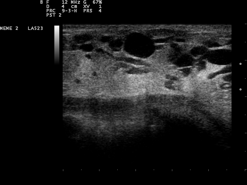
Multiple anechoic cysts in the breast, a cluster of clean black ovals. Simple cysts read as benign, a BI-RADS 2 that needs no biopsy. Image: Nevit Dilmen, CC BY-SA 3.0, Wikimedia Commons.
A handful of benign nodules make up the bulk of what a breast scan finds. The fibroadenoma comes first. It is a solid benign tumor, common in younger women. On ultrasound it reads as an oval, circumscribed, hypoechoic mass that lies parallel to the skin. Around 91 percent of fibroadenomas read hypoechoic. About 58 percent show a clean, circumscribed margin. It feels soft on elastography. A fibroadenoma is the picture of a benign solid nodule.
The simple cyst is the other common find. It is a fluid sac, with no solid tissue at all. On ultrasound it reads anechoic, a clean black oval, with a thin wall and a bright zone of enhancement behind it. A simple cyst that meets all of these is benign with near-certainty, a finding that needs no follow-up. A complex cyst, with debris inside or a thick wall, gets a closer look and sometimes a needle.
Other benign nodules round out the list. Fat necrosis, from an old injury or surgery, can read in many ways, sometimes oily and clearly benign, sometimes firm enough to mimic a cancer. A lipoma reads as a soft, fatty mass. Pseudoangiomatous stromal hyperplasia, PASH, is a benign overgrowth that reads as a solid mass. Each has a benign story behind it, with fat necrosis the readiest to mimic a cancer on the screen.
A few benign nodules need a closer eye. A giant fibroadenoma grows past five centimeters and can crowd the breast. A phyllodes tumor reads much like a fibroadenoma. It grows faster. A fraction of phyllodes tumors turn malignant, so a fast-growing fibroadenoma-like mass goes to biopsy. An intraductal papilloma sits in a duct and can bring a bloody discharge, benign and watched for that reason. The benign label still leaves room for a needle in these.
The common cancers
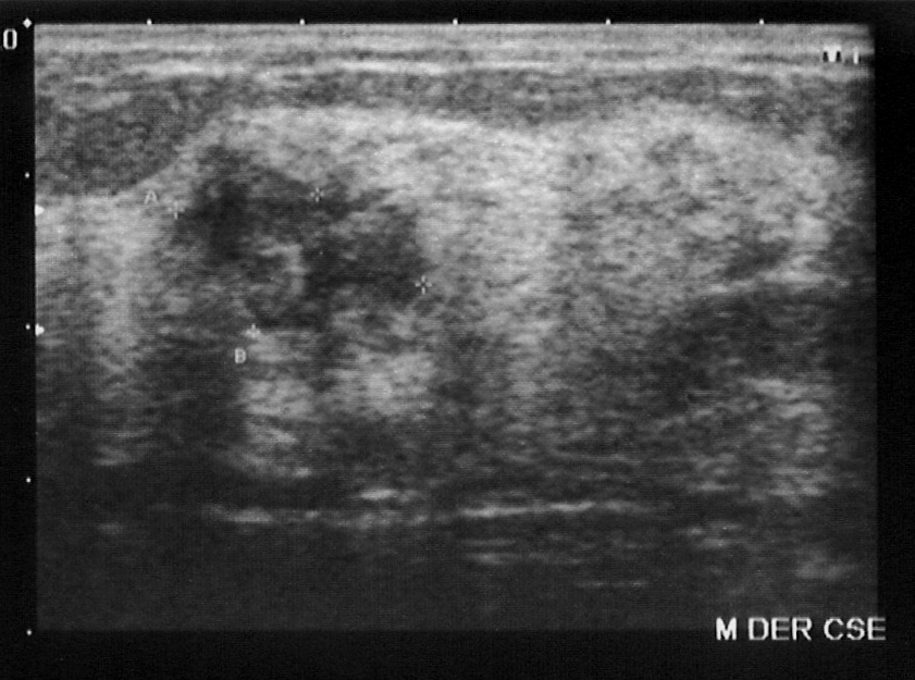
Breast cancer takes a few main forms on ultrasound. Invasive ductal carcinoma is the commonest. It reads as an irregular, hypoechoic mass with a spiculated or angular margin. It often stands taller than it is wide, with shadowing behind it and a bright echogenic halo around it. It distorts the tissue it grows into. Each of these signs raises the suspicion. A mass with several of them is a cancer until a biopsy says otherwise.
Invasive lobular carcinoma is the quiet one. It grows in single-file lines of cells that do not form a clear lump, so it can hide on ultrasound as a subtle patch of shadowing or distortion with no obvious mass. It is the cancer readiest to slip past a scan. A reader who finds shadowing with no mass behind it stays suspicious and looks harder.
Two cancers break the pattern. Mucinous carcinoma and medullary carcinoma can read as round, circumscribed, fairly soft masses, the look of a benign nodule. They are the reason a circumscribed mass is not a guarantee of benign. Ductal carcinoma in situ, DCIS, often shows as calcifications or a change in a duct, with no solid mass at all. The cancers are not all ragged and hard, which is the catch the reader keeps in mind.
Reading benign against malignant
An irregular, hypoechoic breast mass with an unclear margin, measured by calipers. A mass with this disordered look goes to biopsy. Image: SCiardullo, CC BY-SA 3.0, Wikimedia Commons.
The reader reads the benign signs and the malignant signs together on each nodule. The benign picture is one of order: oval, smooth, parallel, soft, with a clean line between nodule and breast. A simple cyst adds the anechoic black and the bright enhancement behind it. A cancer breaks that order: irregular, ragged or spiculated, taller than wide, hard, growing into the tissue with shadowing behind it and a halo around it. The two pictures are clear at their extremes. A textbook fibroadenoma reads benign at a glance, a textbook cancer malignant. The work lies in the middle, where a nodule carries some signs from each side. The reader does not count signs like a tally. One strong malignant sign, a spiculated margin, outweighs several benign ones. A single ragged edge on an otherwise smooth nodule moves it toward biopsy. The reader finds the worst feature and lets it lead. The picture points to the nodule type as well. An oval soft mass in a young woman fits a fibroadenoma, an anechoic mass with enhancement a cyst, an irregular hard mass with shadowing a carcinoma. The reader names the likely nodule, then tests the name against every feature. Stability over time adds to the read. A nodule unchanged across two years of scans behaves like a benign one, even when its look is plain. One that has grown or changed since the last scan moves up the scale, whatever its shape. Age weights the read before the probe touches the skin. A solid nodule in a woman under thirty is far more often a fibroadenoma than a cancer. The same nodule in a woman over fifty carries a higher base risk, so the reader holds a lower threshold for a closer look. A nodule that would be watched at twenty-five goes to biopsy at sixty on the same features. The reader also reads the nodule against the other breast. A matching nodule in the same spot on both sides reads as benign more often than a lone finding. Skin dimpling or a nipple change over a nodule raises the concern sharply. The whole read brings together the look, the stiffness, the flow, the age and the history. A simple cyst needs no second tool of any kind, since its anechoic black and the bright enhancement behind it settle the call on the spot. The behavior over time joins the look on the day.
Elastography, reading the stiffness
Elastography adds a feature the grayscale image cannot show: how stiff the nodule is. It works because cancer tissue runs stiffer than the soft tissue around it. A benign nodule, a fibroadenoma or a cyst, stays soft. A cancer reads hard. The stiffness is the one feature the eye on the grayscale image cannot see.
Two forms are in use. Strain elastography presses the probe lightly and reads how much the tissue deforms, a qualitative soft-to-hard scale shown in color over the grayscale image. Shear wave elastography sends a pulse and measures the speed it travels through the tissue, a number in kilopascals that puts a value on the stiffness. The harder the tissue, the faster the wave runs through it.
Elastography adds specificity to the grayscale read. A soft nodule with a benign look reads benign with more confidence. A hard nodule with a worrying look reads as a cancer with more confidence. The tool does not replace the grayscale features. It adds evidence that settles a borderline call one way or the other, and it spares some benign nodules an unneeded biopsy.
The stiffness can be scored. Strain elastography uses a five-point scale, from a soft nodule that deforms freely to a hard one that holds its shape, with the higher scores pointing to cancer. Shear wave reports a number in kilopascals, where the higher values point to a stiffer, more worrying nodule. The scores turn a feel into a value the next reader can check.
Color Doppler and the blood supply
Color Doppler shows the blood flow in and around a nodule. A cancer grows its own vessels to feed itself, so it often carries more flow than a benign nodule, with vessels reaching into the mass in a disordered pattern. A benign nodule tends to carry little internal flow, or a thin vessel at its edge.
The sign supports the read without deciding it on its own. Some benign nodules, an inflamed one or a fibroadenoma in a young woman, carry brisk flow. Some cancers carry little. Color Doppler adds to a read built on shape and margin, more than it stands alone. A nodule with a worrying shape and heavy chaotic flow reads worse than the same shape with no flow.
Flow also marks the living part of a complex mass. In a part-cystic, part-solid nodule, the solid part with flow inside it is the part to sample, since flowing tissue is living tissue. The probe finds the vessel and aims the needle to where the cells are. A needle into the dead cystic part returns nothing useful.
The pattern of the flow matters more than the amount. A few vessels entering a mass at odd angles, branching inside it, fit a cancer growing its own disordered supply. A single smooth vessel curving around the edge fits a benign nodule. The reader reads the arrangement of the flow, more than the brightness of the color.
Flow helps the read without deciding it.
The traps in the benign-malignant call
The call has known traps, where a nodule reads as the wrong thing. Some cancers look benign. Mucinous and medullary carcinomas can be round, circumscribed and soft, the picture of a fibroadenoma. A circumscribed margin lowers the suspicion. It never clears a mass on its own, since these cancers can carry the benign look. A new round mass in an older woman gets a closer read for this reason.
Some benign nodules look malignant. Fat necrosis can be irregular and hard with shadowing, the picture of a cancer. A history of injury or surgery at the spot points to the benign cause. A complex fibroadenoma can lose its clean margin and read as suspicious. The benign nodule that mimics a cancer goes to biopsy when the history cannot settle it.
The traps are the reason a single feature never makes the call. The reader weighs the whole picture: the look, the stiffness, the flow, the history. Some nodules still need a needle to settle. A biopsy is the right next step when the picture stays unclear, since a missed cancer costs far more than a benign biopsy. The aim is to sample the few that need it and spare the many that do not.
A radial scar deserves its own caution. It distorts the tissue into a spiculated center, the look of a cancer. It can hold a small cancer at its core. A spiculated mass that the history cannot explain goes to biopsy, radial scar or not. The reader treats a spiculated look as malignant until the tissue proves otherwise.
The traps go in both directions. Some benign lesions look hard, some cancers soft.
Settling the call on a handheld unit
A handheld unit makes the benign-malignant read at the bedside, with a high-frequency linear probe. It shows the shape, the margin and the posterior features, adds color Doppler for the flow, and runs elastography where the machine supports it. A nodule that stays unclear after all of it goes to biopsy.
Common questions about benign and malignant breast nodules
How does ultrasound tell a benign breast nodule from a cancer?
It reads the shape, margin, orientation and posterior features, then weighs them with the stiffness on elastography and the flow on color Doppler. A benign nodule tends to be oval, smooth and soft. A cancer reads irregular, ragged and hard.
What does a fibroadenoma look like on ultrasound?
A fibroadenoma is an oval, circumscribed, hypoechoic mass that lies parallel to the skin and feels soft on elastography. Around 90 percent read hypoechoic. It is the common benign solid nodule, usually in younger women.
Can a breast cancer look benign on ultrasound?
Yes. Mucinous and medullary carcinomas can be round, circumscribed and soft, like a fibroadenoma. A circumscribed margin lowers the suspicion without clearing a mass. A new round mass in an older woman gets a closer read.
What is breast elastography?
It measures how stiff a nodule is, since cancer tissue runs stiffer than benign tissue. Strain elastography gives a soft-to-hard color scale. Shear wave elastography gives a stiffness value in kilopascals. It adds confidence to the grayscale read.
Does color Doppler diagnose breast cancer?
No. It shows the blood flow, which supports the read without deciding it. A cancer often carries heavy chaotic flow. Some benign nodules also carry brisk flow. Doppler is weighed with the shape and margin, never alone.
Can a handheld ultrasound tell benign from malignant?
It reads the same shape, margin and posterior features as a cart system, with color Doppler and elastography where supported. A nodule that stays unclear goes to biopsy. The handheld form brings the read to the bedside.
About the Author
Julien Mercier
Senior R&D Engineer · Medical Ultrasound Transducer Development
Senior R&D Engineer with an M.S. in Applied Physics and over 15 years of experience in medical ultrasound transducer development, specializing in the design verification and performance testing of high-frequency imaging transducers. Currently leading the development and verification of the company’s next-generation high-frequency linear-array transducer, responsible for imaging performance evaluation and reliability analysis in preclinical testing. Brings extensive hands-on experience in piezoelectric element tuning, beamforming parameter optimization, and system-level performance testing.