Parathyroid Ultrasound Scanning Handheld Linear Probe
A normal parathyroid gland is too small to find on ultrasound, and that is the point: when one shows up clearly, it has grown. The four glands sit behind the thyroid and set the calcium level in the blood. When one swells into an adenoma, almost always benign, it drives up the blood calcium and the parathyroid hormone, the picture of primary hyperparathyroidism. That enlarged gland is what the scan looks for, told apart from the thyroid and the nodes around it.
Why the parathyroids get scanned
The trigger is a blood result. A high calcium together with a high parathyroid hormone level points to primary hyperparathyroidism. The cause, in the large majority, is a single adenoma, a benign overgrowth of one of the four glands. The cure is surgery to remove the one gland that has grown.
The scan does not make the diagnosis. The bloodwork does. The scan does the next job. It finds the overgrown gland and marks where it sits, so the surgeon can take it out through a small incision, with no wide exploration of the neck.
A focused operation depends on a confident location. A scan that points to one upper-left gland lets the surgeon open over that spot, check the result with a hormone test on the table, and close within the hour. A hormone level that falls after the gland comes out confirms the right target before the patient leaves the room.
Primary hyperparathyroidism is common, more so in older women. Many cases turn up by chance on a calcium test run for another reason. The early symptoms are quiet, a mix of tiredness, kidney stones and thinning bone, so the blood test is usually the real starting point. By the time the scan is ordered, the diagnosis is already in hand and the question is only where the gland sits.
The scan shrinks the operation that follows. A confirmed single adenoma can come out through a two-centimeter cut under local or light sedation. Without a location, the surgeon opens the neck and checks all four glands, a longer case with more to recover from. The few minutes of scanning pay off in a smaller incision and a faster recovery. A clear location also lets the team avoid a wider dissection and the risks near it, the nerve to the voice box and the other parathyroid glands close by.
What a parathyroid adenoma looks like
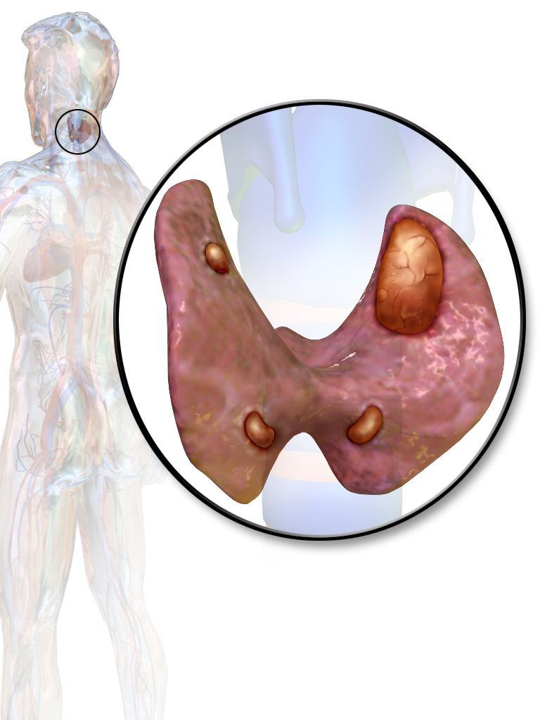
The parathyroid glands sit behind the thyroid. An enlarged one, a parathyroid adenoma, is the gland the scan sets out to find. Illustration: BruceBlaus, CC BY 3.0, Wikimedia Commons.
A parathyroid adenoma reads as a dark, oval mass behind the thyroid. It is homogeneously hypoechoic, darker and more uniform than the thyroid tissue in front of it. A thin bright line, a fat plane, often separates the two. The shape is oval or bean-like, with its long axis running up and down the neck.
Size brings the gland into view. A normal parathyroid measures a few millimeters and stays invisible. An adenoma grows to a centimeter or more, which is what the scan can catch. A large adenoma can reach several centimeters and take on lobules along the way.
The bigger glands are the easier finds. A larger adenoma makes more hormone and shows up more readily on the scan. A small adenoma, making barely enough hormone to raise the calcium, can stay under the probe and force the workup onto the other tests.
The darkness is the first clue. The gland is more cellular than fatty, so it returns little echo and reads almost black against the grey thyroid. A cystic adenoma can hold a fluid pocket inside it. A long-standing one can carry a few flecks of calcium. The uniform dark look, set behind the thyroid and apart from it, is the grayscale signature the reader looks for first.
Not every dark mass behind the thyroid is an adenoma. A thyroid nodule pushing back from the gland can look the part. A small lymph node can read dark and oval. The grayscale look starts the case. The color study and the bloodwork close it.
The polar feeding vessel
The strongest sign on color Doppler is the feeding vessel. A parathyroid adenoma takes its blood from one artery, usually a branch of the inferior thyroid artery, that enters the gland at one end. This polar vessel reaches the upper or lower pole of the oval mass. It avoids the middle. Color Doppler shows it as a stalk of flow arriving at the gland from one side. A polar feeding vessel appears in around 83 percent of parathyroid adenomas, which makes it a dependable marker. The artery often curves around the surface of the gland before it enters, a pattern some readers call a vascular arc. Inside, the adenoma can show a rim of flow or a scattered internal pattern. The polar vessel does more than confirm a parathyroid. It separates one from a lymph node. A lymph node takes its blood through a central hilum, so the flow enters the middle and spreads out. A parathyroid is fed at one pole, with the flow arriving at the end of the gland. The two patterns are distinct enough to tell the structures apart on a clean color study. The vessel also helps find a faint adenoma. A reader who spots a polar artery reaching toward the thyroid can follow it to the gland it feeds, even when the gland is small or set deep behind the thyroid. Power Doppler reads the slow flow of the vessel better than standard color, which helps in a deep or small adenoma. The pattern is not foolproof. A vascular thyroid nodule at the back of the gland can carry its own flow and look the same. A lymph node with a stretched hilum can read the same way. The reader weighs the polar vessel with the shape, the fat plane and the bloodwork before settling the call. A single clean polar vessel counts for more than a busy internal flow. Some adenomas show little color inside at all, so the pole entry stays the steady sign. Power Doppler with a low-flow setting brings out a faint vessel that standard color can miss. The vessel can sit too small to catch on a first pass, so the reader steps up the gain and slows the sweep to bring it out. A clean polar vessel into an oval dark mass behind the thyroid, in a patient with a high calcium, is as close to certain as ultrasound reaches here.
Where to look in the neck
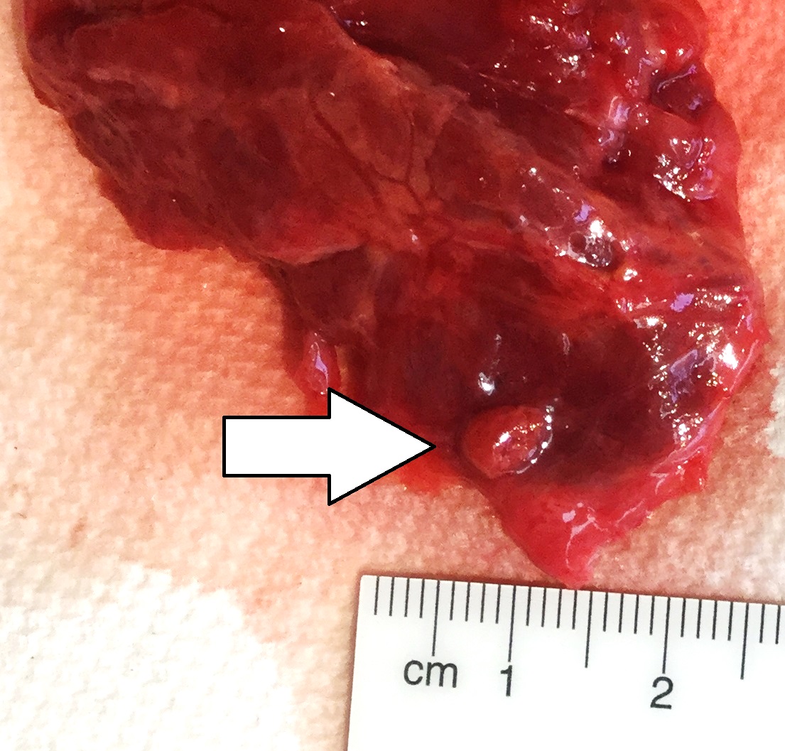
A resected parathyroid adenoma, at the arrow, beside a centimeter scale. The small oval gland is the shape and size the scan looks for behind the thyroid. Specimen: Mikael Haggstrom, CC0, Wikimedia Commons.
A parathyroid adenoma hides in a predictable place. The classic spot sits behind the thyroid, in front of the longus colli muscle along the back of the neck, toward the midline from the carotid artery. The upper pair sits high behind the thyroid, the lower pair near the lower pole or a little below it. The split comes from how the glands form in the embryo and travel down the neck during development.
Swallowing helps the search. A sip of water slides the thyroid and any gland behind it up and down, which carries a hidden adenoma into a clearer plane. Scanning in both the transverse and the long axis covers the area from two directions. A slow, careful sweep behind each lobe finds the adenomas that lie in the neck at all. Tilting the probe and angling under the lower pole of the thyroid reaches a gland that sits a little out of the straight view.
Some glands sit off the map. An ectopic parathyroid can lie in the thymus, low in the chest, in the tracheoesophageal groove behind the gland, or inside the thyroid itself. Reported ectopic sites run to the thymus in around 38 percent, the retroesophageal region in 31 percent, and inside the thyroid in 18 percent. An adenoma the neck scan cannot reach is the common reason ultrasound misses one. A normal neck scan in a patient with clear bloodwork does not rule out an adenoma. It moves the search to the chest.
The embryo explains the hiding places. The lower glands come down with the thymus during development, so a missed lower gland often sits in the chest with thymic tissue. The upper glands travel a shorter path, so they stay closer to home behind the thyroid. Knowing the route tells the surgeon where to look next when the neck comes up empty.
Telling it from the thyroid and the nodes
The hardest call is a parathyroid against a thyroid nodule. A nodule at the back of the thyroid can bulge outward and look like a separate gland. The fat plane settles many of these. A parathyroid sits outside the thyroid capsule, with a thin bright line between the two. A nodule sits inside the capsule, with thyroid tissue wrapped around it.
The fat plane is the first thing to check.
An intrathyroidal parathyroid breaks that rule. It sits inside the gland, with no fat plane to mark it off. A dark, round spot inside the thyroid of a patient with high calcium has to be read as a possible parathyroid, since a missed one inside the thyroid is a known trap. A needle sample washed for parathyroid hormone can settle a doubtful case, since a parathyroid washing returns a hormone level many times the blood reading.
Lymph nodes are the other lookalike. A node behind the thyroid can read dark and oval. The hilum and its central flow set it apart from the polar vessel of a parathyroid. A node also tends to keep a bright fatty center, which a parathyroid does not show. The blood supply is the cleanest way to part the two.
Color Doppler settles many of these calls. A polar vessel into an extrathyroidal oval mass, in a patient with high calcium, marks a parathyroid. A node carries a central hilar vessel. A nodule keeps its vessel inside the thyroid tissue. The blood supply, paired with the bloodwork, settles the structure more often than the grayscale alone.
What ultrasound can and cannot do
Ultrasound finds a single adenoma well, when the adenoma sits in the neck. Reported sensitivity runs from around 55 to 87 percent, and the number depends heavily on the operator and the machine. A skilled reader with a high-frequency probe sits at the top of that range. Poor settings and a quick look drop the result toward the bottom.
The smaller operation is the whole point. A confident neck location lets the surgeon plan a one-gland approach through a single small cut, with a hormone check on the table.
The weak spots are known. A small adenoma can stay below the scan. An ectopic one in the chest sits out of reach of a neck probe. Multigland disease, where more than one gland has grown, is the hardest of all, since the scan can catch one and miss the others. A scan that finds a single adenoma in a patient who in fact has four overgrown glands sets up a failed operation.
Other tests cover the gaps. A sestamibi nuclear scan reaches the chest and finds ectopic glands, with a sensitivity around 60 to 90 percent. A 4D-CT scan reaches about 91 percent and reads multigland disease better. Ultrasound paired with sestamibi raises the yield, from 78 to 88 percent for a single adenoma. The neck scan is the first and cheapest step, with the others held for the cases it cannot solve on its own.
Ultrasound has a second role after surgery. A patient whose calcium climbs again, months or years later, gets a fresh scan to look for a gland that was missed or a new one that has grown. The neck is harder to read after an operation, with scar in the way, so the polar vessel and the bloodwork matter even more the second time. A scan before a repeat operation keeps the surgeon from opening scar tissue blind, which lowers the risk to the voice nerve.
Scanning technique on a handheld unit
A handheld unit scans the parathyroids with a high-frequency linear probe, the same one used for the thyroid. The reader sweeps behind the thyroid in transverse and long axis, adds a color box to find the polar vessel, and uses a swallow to shift a hidden gland into view. The study takes a few minutes at the bedside.
Common questions about parathyroid ultrasound
What does a parathyroid adenoma look like on ultrasound?
It reads as a dark, oval mass behind the thyroid, homogeneously hypoechoic and more uniform than the thyroid in front of it. A thin bright fat plane often separates the two. Adenomas usually measure a centimeter or more.
What is the polar feeding vessel?
It is an artery that enters the adenoma at one pole, usually a branch of the inferior thyroid artery. It appears in around 83 percent of adenomas on color Doppler. The pole entry separates a parathyroid from a lymph node, which takes its blood at a central hilum.
How do you tell a parathyroid from a thyroid nodule?
A parathyroid sits outside the thyroid capsule, with a fat plane between them. A nodule sits inside the gland. The polar feeding vessel and the high blood calcium point to a parathyroid. A needle wash for parathyroid hormone settles a doubtful case.
How accurate is ultrasound for parathyroid adenomas?
Reported sensitivity runs from about 55 to 87 percent, and it depends heavily on the operator. Ultrasound does well with a single neck adenoma. It struggles with ectopic glands and multigland disease, where a sestamibi or 4D-CT scan adds reach.
Where can a parathyroid adenoma hide?
Beyond the usual spot behind the thyroid, an adenoma can lie in the thymus, low in the chest, behind the esophagus, or inside the thyroid itself. A normal neck scan with high calcium moves the search to the chest.
Can a handheld ultrasound scan the parathyroids?
Yes, with a high-frequency linear probe. The reader sweeps behind the thyroid in two planes, adds color Doppler to find the polar vessel, and uses a swallow to bring a hidden gland into view.
About the Author
Julien Mercier
Senior R&D Engineer · Medical Ultrasound Transducer Development
Senior R&D Engineer with an M.S. in Applied Physics and over 15 years of experience in medical ultrasound transducer development, specializing in the design verification and performance testing of high-frequency imaging transducers. Currently leading the development and verification of the company’s next-generation high-frequency linear-array transducer, responsible for imaging performance evaluation and reliability analysis in preclinical testing. Brings extensive hands-on experience in piezoelectric element tuning, beamforming parameter optimization, and system-level performance testing.