Hashimoto Thyroiditis Ultrasound Features Handheld Diagnosis
Hashimoto thyroiditis shows up across the entire gland at once, a diffuse change with no single edge to point to. The body’s own antibodies attack the thyroid. The gland turns dark and uneven on ultrasound, broken into small patches by bands of scar. Reading it means recognizing that spread-out pattern first, then looking for the nodules that can hide inside it.
What Hashimoto thyroiditis is
Hashimoto thyroiditis is the commonest cause of an underactive thyroid in places with enough iodine. The immune system makes antibodies against thyroid proteins. Lymphocytes move into the gland and slowly replace working tissue with scar. The gland can swell early in the disease. It often shrinks over years as the scarring takes hold.
The disease favors women by a wide margin, several times more often than men. It tends to appear in middle age. It clusters with other autoimmune conditions, so a patient with type 1 diabetes or vitiligo carries a higher chance of it. A family history is common.
Two blood tests anchor the diagnosis. Antibodies against thyroid peroxidase, the TPO antibodies, are raised in the large majority of cases. Thyroglobulin antibodies are raised in many. A thyroid hormone panel shows where the gland sits, from normal early on to frankly underactive later.
Iodine has a role here. In iodine-rich diets Hashimoto disease is the common cause of an underactive gland.
The hormone result sets the clinical stage. A normal result with raised antibodies is subclinical disease, watched over time. A low result with a high TSH is overt hypothyroidism, treated with replacement. A related form, postpartum thyroiditis, can flare in the months after a birth and settle on its own.
The change on the screen is diffuse. It runs through the whole gland, both lobes together. A nodule has a discrete edge. Hashimoto disease has none. Ultrasound adds the picture the bloods cannot give. It shows how far the disease has gone and whether a nodule sits among the diffuse change.
The diffuse pattern on the screen
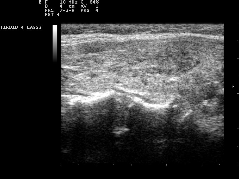
A Hashimoto gland on ultrasound, diffusely dark and coarse. The texture is broken into patches by brighter bands of fibrous scar, the look that covers both lobes. Image: Nevit Dilmen, CC BY-SA 3.0, Wikimedia Commons.
The first sign is darkness. A normal thyroid reads brighter than the strap muscles in front of it. A Hashimoto gland drops to the same level or below, since the lymphocytes that fill it scatter sound less than normal follicles do.
The texture turns coarse. A normal gland reads fine and even. A Hashimoto gland reads patchy, with darker areas broken up by brighter strands. The brighter strands are bands of fibrous scar that run between the patches.
The bright bands give the gland a lobulated, almost cobblestone surface. Some readers call the look a giraffe pattern, after the dark patches set off by pale lines. The pattern covers both lobes, which separates a diffuse disease from a single dark nodule.
A diffusely dark gland is not unique to Hashimoto disease. Graves disease also darkens the gland. A Graves gland tends to read more even, without the patchy scar bands. The patchy, banded texture points to Hashimoto. A smooth dark gland with heavy flow throughout points to Graves.
A few normal findings can mimic the early pattern. A scattering of tiny colloid cysts, each with a comet-tail spark, can look like micronodules until the comet tails give them away. A gland that reads a little coarse, with normal flow and normal antibodies, may sit within normal limits. The diffuse darkness of true Hashimoto disease runs deeper and covers more of the gland.
Micronodules and pseudonodules
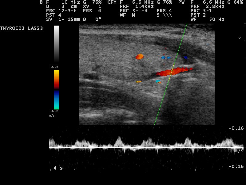
Color Doppler over a Hashimoto gland. The flow runs in the tissue between the dark patches, which carry none of their own. A patch with no internal flow of its own is a pseudonodule, the lymphocyte patch that mimics a nodule. Image: Nevit Dilmen, CC BY-SA 3.0, Wikimedia Commons.
The patches in a Hashimoto gland can read like tiny nodules. These are micronodules, small dark spots one to several millimeters across, scattered through the gland. They are a strong marker of the disease. A gland full of them carries a positive predictive value near 95 percent for Hashimoto thyroiditis on its own. The micronodules are not true tumors. Each one is a patch of lymphocytes ringed by a thin line of scar. The ring is what gives the spot its edge on screen. A true nodule and a micronodule can look alike on grayscale, since both are dark and rounded. The difference shows on color Doppler. A pseudonodule, the lymphocyte patch, carries no internal flow of its own. The blood runs in the normal tissue and the fibrous lines around it. It stays out of the dark patch. This is the giraffe pattern read with color: dark avascular patches set in a mesh of flowing tissue. A real nodule often carries its own internal vessels. So a dark spot with flow inside it draws a second look. A dark spot with flow only around it fits the pattern of the disease. The handheld reader uses the same trick. Drop a color box over a suspicious patch. Flow inside the patch suggests a real nodule that may need sampling. No flow inside, with flow in the bands around it, fits a pseudonodule of Hashimoto. The reading spares a needle in many cases, since a pseudonodule needs no biopsy. The trick has a name in the literature, the giraffe pattern. It rests on one fact: lymphocyte patches do not grow their own blood supply. A reader who learns it stops calling every dark patch a nodule. The handheld screen shows the flow as clearly as a cart system, with the gain set to pick up slow flow. A pseudonodule also lacks the clean round border of a true nodule. Its edge is a ragged scar line, far from a smooth capsule. Micronodules usually measure two to six millimeters across. A true nodule that needs tracking runs larger and stands apart from the even scatter of pseudonodules. A low-flow Doppler preset picks up the faint vessels of a real nodule that a standard setting can miss. The count and spread of micronodules track the disease. A few scattered spots mark an early or mild case. A gland packed with them, edge to edge, marks an advanced one.
Blood flow in Hashimoto disease
Color Doppler sorts Hashimoto glands into a few flow patterns. Early or active disease can carry heavy flow throughout the gland. This heavy diffuse flow, sometimes called a thyroid inferno, overlaps with the look of Graves disease and marks the inflammation. Some readers grade the flow on a simple scale, from a quiet gland to a gland full of color. The grade tracks the activity of the disease at the moment of the scan.
Late or burnt-out disease carries little flow. The scarred gland reads hypovascular. Low flow shows up in close to half of focal Hashimoto cases, against around a fifth of other benign nodules and malignant ones. The drop in flow follows the loss of working tissue to scar.
One pattern carries its own name. A focal patch of intense flow inside a hypoechoic area, a focal thyroid inferno, is specific for a focal form of Hashimoto disease. A solid dark nodule that looks suspicious on grayscale, paired with that focal inferno of flow, often turns out to be a Hashimoto patch. Reading it spares a biopsy that grayscale alone would have prompted.
Power Doppler reads the slow flow of a quiet gland better than standard color, which helps in late disease where the vessels are sparse. Treatment can change the picture. A gland settled on replacement therapy can quiet its flow over months. The grayscale scarring stays. The scan tracks the inflammation. It does not show a cure, only the state of the gland on the day of the scan.
Flow follows the phase of the disease.
How the gland changes in size
Hashimoto disease moves through stages. The gland size moves with each one. Early disease often enlarges the gland. The lymphocytes pack into the tissue and swell both lobes, sometimes enough to feel as a firm, rubbery goiter under the hand. The patient may notice the swelling or feel nothing at all, since the disease is often silent.
Later disease shrinks the gland. As scar replaces working follicles, the lobes grow small and stiff. A long-standing Hashimoto gland can read as a thin, dark, irregular strip of tissue, hard to find against the neck. The isthmus, the bridge between the lobes, can thicken early as a clue.
In children and teenagers, Hashimoto disease is a common cause of a goiter. The gland reads coarse and dark the same way it does in adults. The isthmus often measures thicker than the usual two to three millimeters early on. A gland that grows firm and bumpy, with the diffuse pattern behind it, fits the disease at any age.
Size on its own does not date the disease. An enlarged gland with diffuse change marks an active phase. A shrunken one marks a late phase. The pairing of size with echotexture and flow places the gland on its course. A painful, tender gland that turns dark over weeks points away from Hashimoto, toward subacute thyroiditis, which clears on its own. Hashimoto disease is painless and stays.
How the scan fits with the bloodwork
Ultrasound does not diagnose Hashimoto disease on its own. It works with the bloodwork. Raised TPO antibodies, a diffuse hypoechoic gland and a rising TSH together build the diagnosis. Any one alone leaves room for doubt.
The scan adds what the bloods miss. Antibodies say the immune system is active. They do not say how much gland is left. Ultrasound shows the tissue itself: the scarring, the size and the flow. High antibodies with a near-normal scan suggest early disease in a gland with little damage so far. A shrunken gland with low flow has reached the late stage, whatever the antibody number reads.
The scan also flags trouble the bloods stay silent on. A discrete nodule growing in a Hashimoto gland is the one finding antibodies cannot explain. That nodule gets read on its own merits, since Hashimoto disease and thyroid cancer can sit in the same gland.
The antibody level does not track the gland size, so a high titre does not mean a small gland. TPO antibodies are the more sensitive of the two markers. A patient with stable disease needs no set scan interval. A new lump, a faster change in size, or a pressure symptom brings the patient back to the probe. The scan serves as the tool that watches the tissue over years, scan after scan.
Nodules inside a Hashimoto gland
A Hashimoto gland is busy on the screen, which makes a true nodule harder to spot. The diffuse dark texture hides a dark nodule. The micronodules crowd the field. A reader has to separate a real, discrete nodule from the background of pseudonodules.
A few features mark a real nodule. It has a clear border the pseudonodules lack. It can distort the gland outline or push on a neighbor. Internal flow on color Doppler sets it apart. Microcalcifications, which pseudonodules never show, settle the question. Any of these marks a patch as a nodule that gets scored on its own merits.
The risk is real. Thyroid cancer, chiefly papillary, can grow in a Hashimoto gland at a rate at least as high as in a normal one. Hashimoto disease also carries a small, specific risk of primary thyroid lymphoma. Lymphoma reads as a large, strikingly dark mass, often with bright lines through it and sound passing easily behind it. It grows over weeks in an older patient with long-standing disease. That speed marks it apart from the slow course of the gland itself. A nodule that grows quickly in a known Hashimoto patient gets urgent attention for that reason.
Lymphoma needs a tissue diagnosis fast, often a core biopsy with flow cytology, since a fine needle alone can miss the type. Sampling a nodule in a Hashimoto gland also carries a known catch. The inflamed background can return atypical cells that read as suspicious on cytology, which raises the rate of unclear results. The reader weighs that against the grayscale and the flow before calling a patch in an inflamed gland a cancer.
Reading Hashimoto disease on a handheld unit
A handheld probe shows the Hashimoto pattern as well as a cart system, with a high-frequency linear transducer. The dark, coarse texture and the micronodules stand out on the grayscale image. A quick color sweep separates pseudonodules from real ones. The whole study takes a few minutes at the bedside.
Common questions about Hashimoto thyroiditis on ultrasound
What does Hashimoto thyroiditis look like on ultrasound?
The whole gland turns dark and coarse, broken into patches by bright bands of scar. Small dark micronodules scatter through it. Early disease enlarges the gland, and late disease shrinks it. The change covers both lobes rather than a single spot.
What is a micronodule in Hashimoto disease?
It is a small dark spot, one to several millimeters across, made of a patch of lymphocytes ringed by scar. A gland full of micronodules points strongly to Hashimoto disease, with a positive predictive value near 95 percent.
How do you tell a pseudonodule from a real nodule?
Color Doppler is the key. A pseudonodule carries no flow inside it, with the blood running in the tissue around it. A real nodule often carries its own internal flow, a clear border, or microcalcifications.
Does Hashimoto disease change the size of the thyroid?
Yes. Early disease often enlarges the gland into a firm goiter. Late disease shrinks it as scar replaces working tissue, leaving a small, stiff, irregular gland.
Can you have cancer in a Hashimoto gland?
Yes. Papillary cancer can grow in a Hashimoto gland, and the disease also carries a small risk of thyroid lymphoma. A discrete nodule with a clear border, internal flow, or microcalcifications gets read on its own.
Can a handheld ultrasound diagnose Hashimoto thyroiditis?
It shows the pattern clearly with a high-frequency linear probe. The diagnosis still rests on the scan together with TPO antibodies and a thyroid hormone panel, since no single test settles it alone.
About the Author
Julien Mercier
Senior R&D Engineer · Medical Ultrasound Transducer Development
Senior R&D Engineer with an M.S. in Applied Physics and over 15 years of experience in medical ultrasound transducer development, specializing in the design verification and performance testing of high-frequency imaging transducers. Currently leading the development and verification of the company’s next-generation high-frequency linear-array transducer, responsible for imaging performance evaluation and reliability analysis in preclinical testing. Brings extensive hands-on experience in piezoelectric element tuning, beamforming parameter optimization, and system-level performance testing.