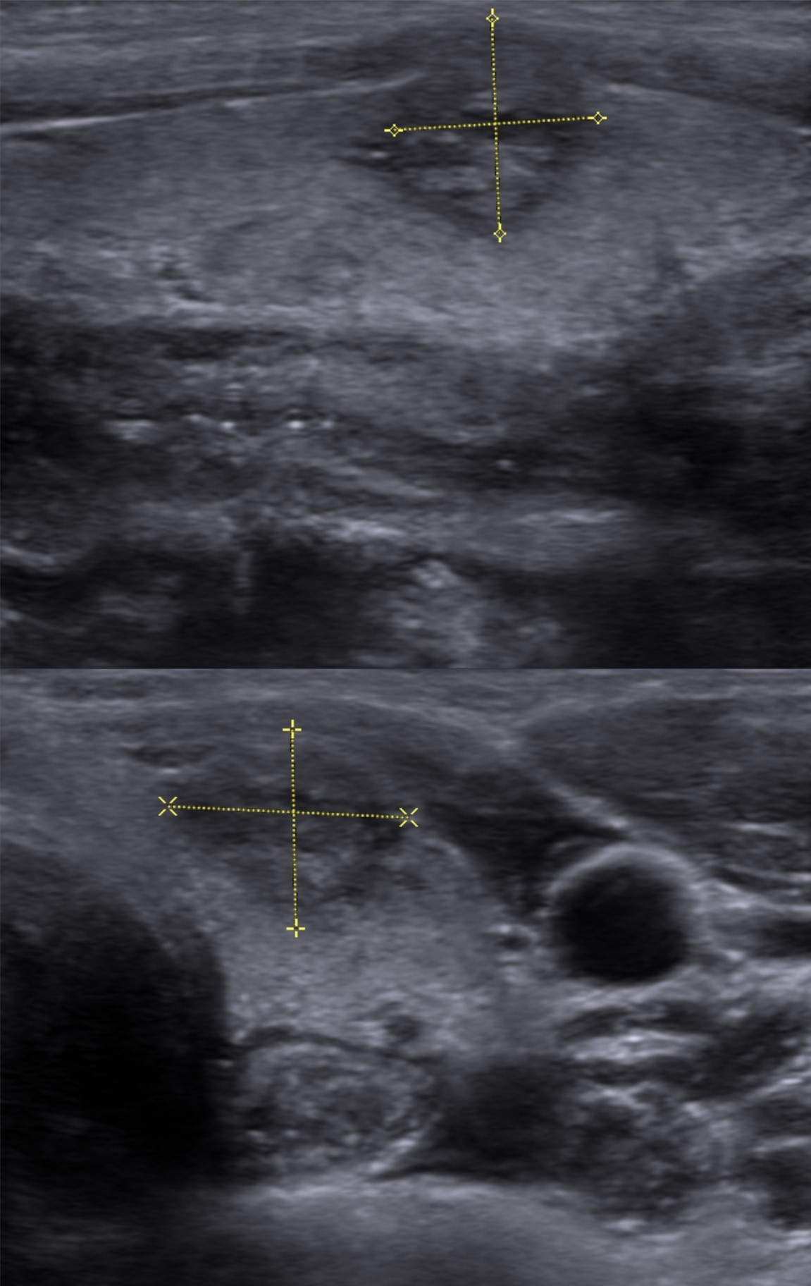
TI RADS Thyroid Imaging Reporting and Data System Handheld Scoring
TI-RADS turns five ultrasound features of a thyroid nodule into a number, and that number, with the nodule size, decides whether it gets a needle, a repeat scan, or nothing further. The American College of Radiology version, published in 2017, scores composition, echogenicity, shape, margin and echogenic foci, then adds the points up. Spotting those features on the screen is one skill; turning them into a defensible total is a separate one.
What the score is built to do
Before TI-RADS, two readers looking at the same nodule could write two reports that pointed two ways. One called a nodule suspicious. Another called the same nodule probably benign. The referring doctor had no shared scale to weigh those words against. TI-RADS was built to close that gap. It fixes a vocabulary, attaches a point value to each finding and ties the final number to a written action.
TI-RADS did not begin with one committee. The Chilean radiologist Horvath proposed an early version in 2009. Kwak published another in 2011. The American College of Radiology consolidated the approach in 2017. That release is the version scored here.
The payoff is measured in needles not taken. Thyroid nodules are common. The large majority are benign. A system that sends every hypoechoic nodule to biopsy buries the clinic in negative results. TI-RADS raises the size bar for the lower levels, so a 2 cm mildly suspicious nodule can be watched, with the needle held back.
The scale also travels. A number written in one clinic means the same thing in the next. A nodule scored TR3 on a handheld unit in a screening camp carries that score to the hospital that receives the referral. A structured report makes the handoff cleaner. The number sits beside the size and the location, so the receiving clinic reads a record where every nodule has the same fields.
The five features and their points
The score is the sum of five feature categories. Each one contributes a fixed number of points. That value never shifts from nodule to nodule.
Composition is read first. A cystic or almost entirely cystic nodule scores zero. A spongiform nodule, built from many tiny cystic spaces, also scores zero. A mixed cystic and solid nodule scores one. A fully solid nodule scores two. Composition alone can place two points on the board before another feature is weighed. The two zero-point compositions are grouped for a reason. A nodule that is largely fluid, or a sponge of tiny cystic spaces, almost never holds cancer, so neither draws a point. Echogenicity is judged against the normal gland and the strap muscles. It covers the widest range of any single category. An anechoic nodule scores zero. A hyperechoic or isoechoic nodule scores one. A hypoechoic nodule, darker than the gland, scores two. A very hypoechoic nodule, darker than the strap muscles, scores three. The very hypoechoic call is the harder one, because the reference shifts from the gland to the darker strap muscle. Shape is read in one transverse plane and has only two outcomes. A nodule wider than it is tall scores zero. A nodule taller than it is wide scores three. The ratio is read where the height and the width sit side by side on a frozen image. That jump is the largest single-feature move in the system, because a taller-than-wide pattern crosses the normal tissue planes of the gland. Margin describes the border between nodule and gland. A smooth margin scores zero. An ill-defined margin, where the edge fades without a sharp line, also scores zero, since a soft edge is common in benign disease. A lobulated or irregular margin scores two. Extrathyroidal extension, where the nodule breaks the capsule, scores three. Margin is the feature readers disagree on, since a blurred edge can read as smooth or ill-defined depending on the gain. Echogenic foci is the final category, read for the brightest type present. None, or a large comet-tail artifact from colloid, scores zero. Macrocalcifications score one. Peripheral or rim calcification scores two. Punctate echogenic foci, the bright specks that often mark psammoma bodies, score three. The gap between those last two values matters. Punctate foci track with papillary cancer. Coarse macrocalcifications are a weaker, less specific sign. The five categories are independent. A single nodule can draw points from every one of them. That independence is how a solid, very hypoechoic, taller-than-wide nodule with an irregular margin and punctate foci reaches a total well above seven, with no one feature deciding the level on its own.
Adding the points into a level
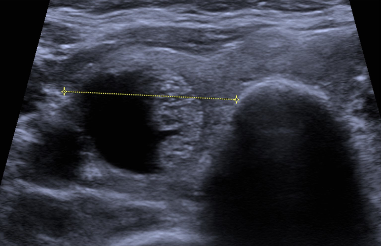
A solid, hypoechoic thyroid cancer measured in two planes. A dark nodule with an unclear margin draws points on echogenicity and margin, which lifts the total toward the high levels. Image: Hellerhoff, CC BY-SA 4.0, Wikimedia Commons.
The five values are summed. The total drops the nodule into one of five levels. A total of zero is TR1, benign. A total of two is TR2, not suspicious. A total of three is TR3, mildly suspicious. A total of four, five, or six is TR4, moderately suspicious. A total of seven or more is TR5, highly suspicious. The levels are not evenly spaced. TR2 holds a single total. TR4 spans three of them. The spacing reflects how points cluster in real nodules.
Each level carries a measured risk of cancer, drawn from large validation series. TR1 runs near 0.3 percent. TR2 sits around 1.5 percent. TR3 is close to 4.8 percent. TR4 reaches about 9.1 percent. TR5 climbs to roughly 35 percent. These percentages come from series of thousands of nodules checked against cytology or surgery, which is what anchors them. The numbers show why the score does not end the workup. A TR5 nodule still has close to a two-in-three chance of being benign, so the level sets a degree of suspicion and stops there.
A worked example shows the sum in action. Take a solid nodule, which is two points. It reads hypoechoic, two more. It is wider than tall, zero. Its margin is lobulated, two more. It holds a single macrocalcification, one. The total is seven, which lands at TR5. Drop the calcification and the lobulation. The same nodule scores four, which is TR4. Two findings moved it a full level. The arithmetic runs at the bedside without a chart.
TR4 carries a large share of nodules in routine practice. It spans three totals, from four to six. That range is where benign and malignant nodules overlap on ultrasound, which is why the level leans on size to decide the next move.
A nodule like the one above reaches TR5 on sight, before the points are tallied.
How size turns a level into a biopsy
A benign macrofollicular nodule with several cystic spaces. A cystic make-up sits at the low-scoring end, where the size bar rarely calls for a needle. Image: Hellerhoff, CC BY-SA 4.0, Wikimedia Commons.
The level alone does not send a nodule to biopsy. Size does that. Each level has its own size bar. A TR5 nodule is sampled at 1 cm and above. A TR4 nodule is sampled at 1.5 cm and above. A TR3 nodule is sampled at 2.5 cm and above. TR1 and TR2 nodules are not sampled on imaging grounds. The bar rises as suspicion falls, so a low-suspicion nodule has to grow large before a needle is justified.
Below each biopsy bar sits a follow-up band. A TR5 nodule from 0.5 to 0.9 cm is watched with repeat ultrasound. A TR4 nodule from 1 to 1.4 cm is watched. A TR3 nodule from 1.5 to 2.4 cm is watched. A watched nodule is rescanned at intervals, often about a year apart. A nodule that stays stable across scans eventually leaves surveillance. A nodule that grows past its size bar moves to biopsy.
One rule overrides the rest. Nothing under 5 mm goes to a needle, even at TR5.
A spongiform nodule shows the scale from its low end. Its composition scores zero. Its total holds at TR1 or TR2. The size bar never comes into play, so a 3 cm spongiform nodule is left alone. A 1 cm solid nodule goes to the needle.
Three systems, three size cutoffs
ACR TI-RADS is not the only system in use. The European Thyroid Association publishes EU-TIRADS. The Korean Society of Thyroid Radiology publishes K-TIRADS. Each one sorts nodules into five levels. Each one ties the level and the size to a biopsy decision. The route to the level is where they part. Three systems exist because guidelines grew up in separate regions. A reader uses whichever one the local protocol names. The features under all three come from the same ultrasound.
ACR TI-RADS reaches the level by adding points across five categories. The reader scores each feature and sums them. A total maps to TR1 through TR5.
EU-TIRADS reaches the level by pattern. The reader matches the whole nodule to a described category. EU-TIRADS 2 is benign at a risk near zero. EU-TIRADS 3 is low risk at 2 to 4 percent. EU-TIRADS 4 is intermediate at 6 to 17 percent. EU-TIRADS 5 is high risk at 26 to 87 percent. The size bars run 20 mm for category 3, 15 mm for category 4 and 10 mm for category 5.
K-TIRADS also works by pattern, with bars of its own. K-TIRADS 2 is benign. Biopsy does not apply. K-TIRADS 3 is low suspicion at 3 to 10 percent, sampled above 2 cm. K-TIRADS 4 is intermediate at 10 to 40 percent, sampled in a 1 to 1.5 cm band. K-TIRADS 5 is high suspicion above 60 percent, sampled above 1 cm.
The systems also weigh single features in their own way. EU-TIRADS treats marked hypoechogenicity as a high-risk sign on its own. ACR TI-RADS folds that finding into the three-point very-hypoechoic score and lets the sum decide. A clinic that switches systems re-reads its criteria along with its cutoffs.
The three land close together at the high end. A clearly suspicious 1 cm nodule meets the bar under all three. They separate at the low end. A 2.2 cm low-suspicion nodule sits below the ACR bar of 2.5 cm. It sits above the EU bar of 20 mm. It sits above the K-TIRADS bar of 2 cm. The same nodule, scored by the book, goes to biopsy under two systems and to follow-up under the third. A clinic picks one system and applies it across every scan.
Scoring on a handheld screen
A handheld unit scores a nodule the same way a cart system does. Two habits protect the number. Freeze and magnify before measuring shape, since a taller-than-wide call turns on a millimeter. Use the highest frequency the probe allows to bring out punctate foci, which fade on a low-frequency image.
Where the score misleads
A score is only as reliable as the features behind it. A few patterns send the score wrong. Spongiform is the first trap. A nodule has to be more than half spongiform by volume to score zero on composition. A nodule with a few cystic spaces inside solid tissue is solid. It scores two. Calling it spongiform by mistake drops the score by two points and can move a nodule out of biopsy range.
Echogenic foci inside a mixed cystic and solid nodule are the second trap. Bright specks against colloid are often comet-tail artifact. They get read as punctate calcification by mistake. Scoring them as punctate foci adds three points the image does not support.
Isoechoic nodules mislead in a third way. A nodule that matches the gland in brightness can hide its own margin. An unread margin slips to a low score by habit. The fix is a wider field and lower gain, which brings the edge out against fat or muscle before the reader scores it.
Size itself can throw the level off. The longest diameter sets the size bar, so an under-measured nodule can fall below its cutoff and miss a needle it should have. Measuring on the plane that shows the nodule at its largest keeps the size honest.
Follow-up adds one more trap, this time in time. The score is read fresh each time. A nodule that was TR3 last year is scored again from the current image. Last year’s number is set aside, since calcification and margin shift as a nodule evolves. The number reflects today’s image and nothing else.
Common questions about TI-RADS scoring
What does a TI-RADS score count?
It counts five ultrasound features of a nodule: composition, echogenicity, shape, margin, and echogenic foci. Each feature is worth a set number of points, from zero to three. The points are added into a single total that sets the risk level.
How do the points become a level?
A total of zero is TR1. Two is TR2. Three is TR3. Four to six is TR4. Seven or more is TR5. The level rises with the total, and each level carries a higher measured risk of cancer, from near 0.3 percent at TR1 to roughly 35 percent at TR5.
At what size is a TR5 nodule sampled?
A TR5 nodule is sampled at 1 cm and above. Between 0.5 and 0.9 cm it is watched with repeat ultrasound. Under 5 mm it gets neither biopsy nor follow-up, even though it sits in the highest level.
How does EU-TIRADS differ from ACR TI-RADS?
ACR TI-RADS reaches a level by adding points across five features. EU-TIRADS reaches a level by matching the nodule to a described pattern. The two also set their own size bars. EU-TIRADS samples a high-risk nodule above 10 mm, close to the ACR 1 cm bar for TR5.
Can TI-RADS be scored on a handheld device?
Yes. The features read the same on a handheld screen. Freezing and magnifying the image before measuring shape, and using the highest available frequency for echogenic foci, keep the score reliable.
Does a high score mean cancer?
No. A high score means high suspicion. It is not a diagnosis. A TR5 nodule carries a cancer risk near 35 percent, so the majority of TR5 nodules turn out benign after biopsy.
About the Author
Julien Mercier
Senior R&D Engineer · Medical Ultrasound Transducer Development
Senior R&D Engineer with an M.S. in Applied Physics and over 15 years of experience in medical ultrasound transducer development, specializing in the design verification and performance testing of high-frequency imaging transducers. Currently leading the development and verification of the company’s next-generation high-frequency linear-array transducer, responsible for imaging performance evaluation and reliability analysis in preclinical testing. Brings extensive hands-on experience in piezoelectric element tuning, beamforming parameter optimization, and system-level performance testing.