


































Probe design
192 versus 128 Element Handheld Ultrasound Clinical Image Quality
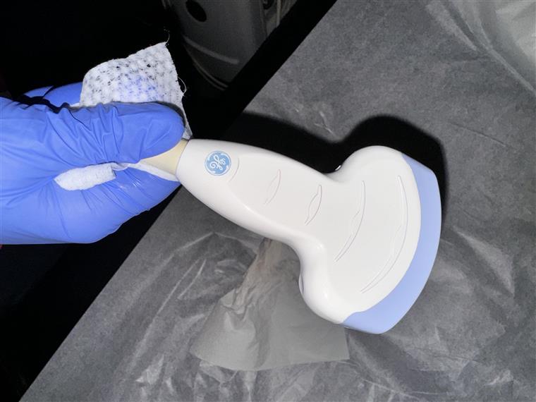
Whether a handheld probe carries 128 elements or 192 shows up less on the spec sheet than in the image itself. The elements are the piezoelectric crystals lined up across the probe face, and their number sets the density of the scan lines, the width of the active aperture and the field of view. More elements fill the picture out, which is part of what separates a cheaper probe from a dearer one.
What the element count sets
Each element sends and receives along one path, and the array fires them in groups to build the image, so the count of crystals across the face decides three things a reader sees on the screen. The first is line density: more elements across a given width lay down more scan lines per centimetre, which sharpens the side-to-side, the lateral, detail. The second is the aperture, the span of elements firing together to form one focused beam, where a wider span draws a tighter beam and holds it through more depth. The third is the field of view, the breadth the image covers at a given line density. Two numbers sit behind the count. The pitch is the spacing between neighbouring crystals, centre to centre, and a small gap between them, the kerf, keeps each one acoustically separate. The array also does not fire every element at once: it fires a sliding sub-aperture and steps it along the face to sweep the lines, widening the group for a deeper focus in a trick called a dynamic aperture. More elements at a fine pitch give the beam-former more to work with, for steering and for focusing alike. The same aperture shapes the side lobes, the weaker echoes a beam throws off to its sides that paint faint clutter into the picture; a wide, well-weighted aperture, with apodization tapering the edge elements, holds those lobes down, and the clutter they would have raised shows most in a space that should read black, like a full bladder or a cyst. None of this reaches the reader as a count of elements. It reaches the screen as line density, as the tightness of the focused beam, and as how clean a dark space reads, which is why two probes of the same count can hand back different pictures once the rest of the imaging engine is weighed in. Pitch carries a second consequence a spec sheet rarely names. Spaced too far apart, the elements let the beam throw grating lobes, false copies of the echo that ghost into the picture, so a fine pitch buys a cleaner field along with a sharper one. The count also says nothing about the third dimension. The crystals line up in a single row, and the beam stays thin across that row. In the plane at right angles, the elevation plane, the beam spreads, held in only by a lens or a second row of elements. A probe that reads crisp on the screen can still average a slice several millimetres thick. That slice thickness, set by the elevation focus the count does not touch, decides how cleanly a small deep structure stands clear of its neighbours. The number is where the picture starts, not where it ends.
What 192 elements give
A 192-element array lays more scan lines across the same field. The lines sit closer together, so the lateral detail is finer and a small structure holds its shape toward the edges of the image. The wider run of elements also forms a broader aperture, which focuses a tighter beam and keeps the focus cleaner across the depth. A vessel wall, a nerve, a fine tendon fibre reads with a crisper edge.
The wider element run carries a wider field of view at full line density. A 192-element linear face covers more breadth without thinning its lines, so a long structure like a vessel running down a limb stays in one frame at full detail. The extra elements feed a heavier stream of channel data, which a capable processor turns into the sharper picture. The count suits the work that lives on fine surface detail, where the difference between a clean wall and a blurred one changes the read.
The count reads against the probe type. A linear array spreads its elements across a wide flat face, where 192 against 128 shows in the breadth of the field and the fineness of the lines. A phased array packs fewer elements into a small footprint for the heart, the count there set by the rib window more than the field. A convex array curves its elements for the abdomen. The same number lays a different line spacing on each face, since the face widths differ.
A wider aperture also gathers more of the returning echo. More elements listening together collect a stronger signal from depth, which lifts the sensitivity on a faint return. Frequency remains the main driver of penetration, so the gain from the broader aperture is modest. It still helps a weak echo read at the far edge of the range.
What 128 elements give
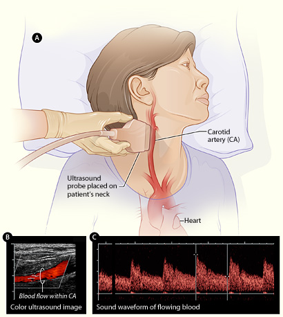
A 128-element array carries the line density a focused point-of-care study works with. The picture holds the detail an everyday question needs.
That picture comes in a smaller, lighter body, on less power and at a lower cost.
Image quality rests on more than the element count
The count is one input among several. Frequency sets the baseline detail: a high-frequency probe draws finer structure than a low one, whatever its element count. The beam-forming, the way the machine times and weights its elements, shapes the focus alongside the raw number of elements. Harmonic imaging cleans a hazy picture by listening at twice the sent frequency. A compound mode blends several beam angles into one steadier frame. The screen and the processor decide how much of the captured detail a reader ever sees. A 128-element probe with strong beam-forming and harmonic imaging can read cleaner than a 192-element probe with weak processing behind it. The honest test is the picture the probe makes on the hardest case the work will meet, read on its own screen, against the structures the clinician scans most.
The element count is a fair first question for a buyer, not the whole answer.
A picture carries three kinds of resolution, and the element count touches only one. The detail along the beam, the axial resolution, rests on frequency and pulse length. Telling one shade of grey from the next, the contrast resolution, leans on the processing. What the count drives is the lateral resolution, the detail across the beam, which sharpens with the denser lines and wider aperture a higher count brings.
Line count trades against frame rate. Each scan line takes time to send and to listen for the echo. More lines per frame, from a higher count at full density, makes each frame take longer, so the frame rate falls. A fast target like a heart valve needs a high frame rate to read without blur. A still target like a tendon can spend the time on lines. A machine holds the balance, narrowing the field or thinning the line density to keep the frame rate up when the motion calls for it.
A dead element shows on the screen as well. A crystal that fails leaves a dropout, a dim line down the image where its scan line should sit. A probe with more elements loses a smaller share of the picture to one dead crystal, since each line carries less of the whole. A drop-out test, scanning a flat surface and watching for a dark stripe, catches a failing element on either count before it costs a read.
Which count suits which work
The fit follows the kind of detail the work lives on. Fine surface work rewards the closer line spacing of a 192-element face. A vascular service reads that detail all day, as does a musculoskeletal clinic placing a needle near a nerve. A fine tendon, a small nerve, a shallow vessel wall reads sharper with the extra lateral detail. These services lean toward the higher count and carry the heavier, costlier probe for the picture it returns.
A focused point-of-care service reads broader questions at depth. An emergency physician checking for free fluid, a critical-care team sizing a heart, a rural clinician sweeping the abdomen reads answers the 128-element count holds well. For this work the lighter probe, the longer battery, and the lower cost match the daily caseload, where the picture the standard count makes answers the question the scan was run to settle. A buyer reads the choice against the structures the work scans most and the hours the probe stays in the hand.
A buyer can settle the count over the structures the work scans. A vascular and nerve list run on a 192-element linear face reads the fine walls and fascicles the work turns on. A mixed point-of-care day, abdomen to heart to a quick line, runs well on a 128-element probe that stays light through the shift. The count follows the picture the work needs, set against the weight and the battery a long day asks for.
Common questions about element count and image quality
What does the element count mean on an ultrasound probe?
It is the number of piezoelectric crystals lined up across the probe face. The crystals fire in groups to form the scan lines that build the image. Handheld probes ship most often with 128 or 192 elements.
How does element count affect image quality?
More elements across the same width lay down denser scan lines, which sharpens the side-to-side detail, and form a wider aperture, which focuses a tighter beam. The count also sets how wide a field the probe covers at full line density.
What does a 192-element probe give?
Finer lateral detail from closer scan lines, a tighter focused beam from a wider aperture, and a wider field of view at full line density. It suits fine surface work like vascular, musculoskeletal, and nerve imaging.
What does a 128-element probe give?
The line density a focused study reads, in a smaller, lighter probe that draws less power, costs less, and runs cooler. It suits point-of-care work across the abdomen, the heart, and the vessels.
Is element count the only thing that sets image quality?
No. Frequency, beam-forming, harmonic imaging, compound imaging, the processor, and the screen all shape the picture. A 128-element probe with strong processing can read cleaner than a 192-element probe with weak processing.
Which element count should a buyer choose?
The one that matches the structures the work scans most. Fine surface detail rewards 192; focused point-of-care questions across depth are well met by 128 at a lighter weight and longer battery life.

About the Author
Julien Mercier
Senior R&D Engineer · Medical Ultrasound Transducer Development
Senior R&D Engineer with an M.S. in Applied Physics and over 15 years of experience in medical ultrasound transducer development, specializing in the design verification and performance testing of high-frequency imaging transducers. Currently leading the development and verification of the company’s next-generation high-frequency linear-array transducer, responsible for imaging performance evaluation and reliability analysis in preclinical testing. Brings extensive hands-on experience in piezoelectric element tuning, beamforming parameter optimization, and system-level performance testing.