


































Probe coverage
3 in 1 Convex Linear Cardiac Probe Wireless Ultrasound Benefits
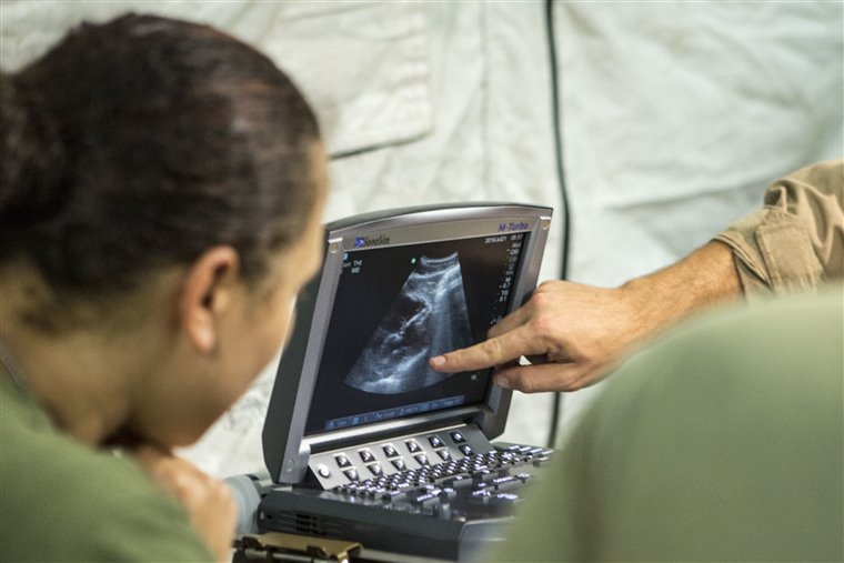
A three-in-one probe is a single handheld transducer that holds three scanning modes in one head: a convex mode for the deep abdomen, a linear mode for the shallow vessels, and a cardiac mode that slips between the ribs to the heart. It answers a wide clinical question from one device, in a single unbroken scan. A sick patient rarely raises one question: chest pain points to the heart, then to the lungs and the great vessels behind them, and a road-traffic victim raises that same wide spread in one sweep. One tool that crosses all three reads a whole sick patient, in the minutes that matter.
What each of the three modes does
The three modes answer three depths. The convex mode runs at 2 to 5 megahertz across a wide curved face, reaching twenty to thirty centimetres into the abdomen for free fluid, the deep organs, a pregnancy. The linear mode runs higher, 5 to 12 megahertz on a flat face, holding detail near a tenth of a millimetre in the first few centimetres, the range a surface vein or the lung lining sits in. The cardiac mode runs low, 1 to 5 megahertz through a footprint small enough to fit between the ribs, where it trades fine resolution for the fast frame rate that freezes a moving heart wall. Together they span 1 to 12 megahertz, a range no fixed probe covers, and the reader turns the depth up for a larger body.
A focused bedside study needs that span. Several emergency protocols ask for these views in sequence, on one patient, in one sitting. A clinician with all three modes runs the sequence without changing tools. A clinician with one mode must stop to fetch a second probe. The patient has to be found again. The reading loses its continuity. The three-in-one design keeps that reading continuous.
The protocols that need all three at once
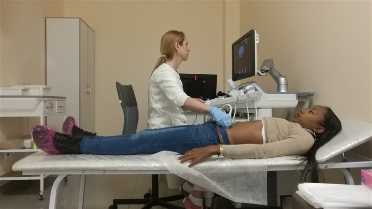
The trauma scan shows the design at work. The eFAST exam checks four abdominal windows plus the chest. The right and left upper quadrant windows show where free blood pools first, between the liver or the spleen and the kidney, the suprapubic window shows the pelvis behind the bladder, and the subxiphoid window shows the sac around the heart. The chest windows then read the lung surface, where a healthy lung slides and a collapsed one stays still. Each of the three modes takes a part of that one scan, the convex reading the abdomen, the cardiac the pericardium, the linear the lung surface, so a three-in-one probe walks the whole protocol without leaving the hand. The shock assessment, the protocol named RUSH, runs the same way, and a trained reader finishes it in under two minutes, reading the heart for its pumping and filling, the inferior vena cava for the circulating volume, the Morison’s pouch for hidden blood, the aorta for an aneurysm, the lungs for a pneumothorax. Clinicians recall those five areas as HI-MAP. Each finding narrows the cause of the shock, and each comes from one mode of the same probe, so a swap between steps would cost a crashing patient minutes and break a sequence that has to be read as one picture. A breathless trauma patient shows the same breadth in miniature, the linear mode reading the lung sliding, the cardiac mode turning up to the chest for fluid or air, both signs caught from the probe that just read the belly and the heart. The modes reach past these named protocols, the convex finding gallstones or an early pregnancy, the linear guiding a vein puncture or hunting a leg clot, the cardiac grading the squeeze and checking the sac for fluid. Behind all three sits a single silicon chip, thousands of micro-machined sensors under one face, with software reshaping the beam for each mode so the reader changes no part of the probe. The chip carries a trade name, CMUT, the capacitive micromachined ultrasound transducer, with a piezoelectric cousin called PMUT, and both pack a whole probe family onto one piece of silicon. The point is not that one probe does three jobs. It is that it does them in one unbroken sweep, where each finding builds on the one before into a single line of reasoning. A swap that sets the probe down drops that thread, and on a crashing patient the thread is the diagnosis itself.
Other named protocols run on the same probe. The BLUE protocol reads the lungs for the cause of breathlessness, checking for lung sliding, for B-lines, for fluid at the base. The FOCUS exam looks at the heart for a pericardial effusion, a weak squeeze, a strained right side. Each protocol leans on one or two of the three modes. The single probe carries them all.
The benefits that reach past the scan
The breadth saves money. One three-in-one probe costs far less than three separate probes. A clinic that cannot afford a full set can afford one device. Ultrasound reaches a budget that three probes would shut out.
Training is simpler. A trainee learns one device. The same handling serves every mode. Three separate probes mean three handling patterns to learn. One body shortens the training.
The probe is lighter. One probe in a pocket weighs less than three in a bag. A ward round, a field call, a one-drawer clinic each needs only the single probe. The work that once filled a cart now fits a pocket. The wireless image reaches past the one reader. A second clinician watches the same screen. A teacher reviews a trainee’s scan as it runs. A specialist reads it from another site over the link.
The imaging modes on one probe
The probe carries more than the grey anatomy picture. B-mode draws that grey picture, the base of every scan. M-mode plots one line against time. It catches fast motion: the lung sliding, a valve opening, a wall thickening through the beat.
Colour Doppler paints flow onto the grey image. One colour marks blood coming toward the probe. Another marks blood moving away. A vessel lights up. A leaking valve shows its jet.
Pulsed-wave Doppler reads the speed of flow at one chosen depth. It measures a stroke distance for a cardiac output. Continuous-wave Doppler reads the top speed along the whole beam. It catches a tight valve jet a gated mode would miss.
A few built-in tools sharpen the picture. Harmonic imaging cleans a hazy abdomen by listening at twice the sent frequency, and speckle reduction smooths the grainy texture left in the raw image. A compound mode steadies the frame by blending several beam angles, and a wide dynamic range holds the bright echoes and the faint ones in one picture. Each loads from the preset, turned on when the picture needs it.
The views each mode reaches
The convex mode opens the abdomen. It reaches the four FAST windows for free fluid, the aorta, the kidneys, a second-trimester pregnancy.
The linear mode works the surface: a vein before a cannula, a leg vein for a clot, the lung sliding under the ribs, a thyroid or a swollen joint.
The cardiac mode reaches the heart through the rib gaps: the four-chamber view from the apex, the parasternal view for the valves, the inferior vena cava for filling, the sac for fluid.
Setting the probe for each scan
A preset loads the settings for each job. The abdomen preset sets a deep field at a low frequency. The vascular preset sets a shallow field at a high one. The cardiac preset speeds the frame rate for the moving heart.
Three controls fix most pictures. Depth sets how far down the screen reaches. Gain sets the overall brightness. The focus marks the depth of the sharpest detail. A reader sets the depth to the target, then trims the gain until the picture reads clean.
What the numbers on the box mean
A buyer reads a few figures first. The probe weighs around three hundred grams. It spans 1 to 12 megahertz across the three modes. It scans out to about thirty centimetres at the deep end. It runs one to two hours on a charge. A regulatory clearance stands behind it, a CE mark in Europe or an FDA clearance in the United States.
The records follow the scan. The clinician freezes a frame, drops a caliper, saves the measurement to the study. A short clip records a beating heart for a second read. The study uploads to the record under the patient’s name.
The learning it takes
The breadth asks for training. One set of controls carries across every mode, so the handling transfers from one to the next. A reader learns one mode at a time, the convex view first, the cardiac last, since the rib windows are narrow and the heart keeps moving.
The features built into the probe
The probe does its own beam-forming inside the head. It sends the image to a phone or tablet over Wi-Fi. No cable runs from the hand to the screen. The clinician scans one-handed. The screen sits propped nearby. The wireless link takes the probe where a cart could never follow.
A lithium battery sits inside the body. A full charge runs about one to two hours of continuous scanning. The probe tops up on a charging pad between sessions. A spare battery covers a long field shift.
Scans save onto the device. A linked cloud account holds a copy. The clinician exports them as standard DICOM files into the patient record. A bedside image reaches the notes with no transfer step.
The sealed body wipes down between patients, with no connector crevice to trap anything.
On-board software measures the routine figures. An ejection fraction traces on its own. A bladder volume reads from one tap. The inferior vena cava gauges the same way. The reader checks each number against the live picture.
Where the design fits best
The design fits the clinician who reads many systems on one patient. The emergency physician is the clearest case. One shift brings a chest pain, a trauma call, a breathless patient, a hard line to place. Each calls for a different mode, and the probe meets them all from one pocket.
Critical care uses it the same way. A team sweeps a deteriorating patient across the heart, the lungs, the abdomen in one round. The probe carries all three with no swap.
A rural clinic often holds one imaging device for a whole district. The three-in-one probe covers the abdomen, the pregnancy, the heart, the lung from that single tool. A field team carries the same reach in a coat pocket.
One encounter shows the flow in practice. A breathless patient arrives. The convex mode opens the abdomen for free fluid. The cardiac mode reads the heart for its squeeze. The linear mode checks the lungs for sliding. The probe stays in the hand across all three. The cause of the breathlessness shows in a few minutes.
Point-of-care work runs on this breadth. A bedside question starts wide open. The probe answers it fast. It moves to the next system on the same patient. One probe reads the whole sick patient in a single sweep.
Common questions about the three-in-one probe
What is a three-in-one ultrasound probe?
It holds three imaging modes in one body: a convex mode for the deep abdomen, a linear mode for shallow structures, a cardiac mode for the heart. One device does the work that once took three separate probes.
What frequency does each mode run at?
The convex mode runs at 2 to 5 megahertz, the linear at 5 to 12, the cardiac at 1 to 5. The set spans 1 to 12 megahertz.
Which clinical protocols can it run?
It runs the eFAST trauma scan, the RUSH shock assessment, the BLUE lung protocol, the FOCUS cardiac exam. Each draws on one or more of the three modes.
How does the probe send its image to a screen?
The probe does its own beam-forming. It sends the image to a phone or tablet over Wi-Fi, with no cable from the hand to the screen.
What can the on-board software measure?
It measures the routine figures: an ejection fraction, a bladder volume, the inferior vena cava. The reader checks each against the live picture.
Who gets the most use from a three-in-one probe?
Emergency physicians, critical-care teams, rural clinicians, field and disaster responders. Anyone who reads several systems on one patient from one pocket.

About the Author
Julien Mercier
Senior R&D Engineer · Medical Ultrasound Transducer Development
Senior R&D Engineer with an M.S. in Applied Physics and over 15 years of experience in medical ultrasound transducer development, specializing in the design verification and performance testing of high-frequency imaging transducers. Currently leading the development and verification of the company’s next-generation high-frequency linear-array transducer, responsible for imaging performance evaluation and reliability analysis in preclinical testing. Brings extensive hands-on experience in piezoelectric element tuning, beamforming parameter optimization, and system-level performance testing.