


































Cardiac function
Right Ventricular Function Assessment Handheld POCUS
The right ventricle gives way faster than the left, and it shows its trouble in shapes the eye catches in seconds. Built thin for the low pressure of the lungs, it dilates and weakens within hours when that pressure climbs, which is what makes it a quick read on a sick chest. A handheld probe asks three plain things at the bedside: whether the chamber is enlarged, whether it still squeezes, and whether a clot has blocked the lungs in front of it. None of the three needs a tracing or a calculation.
Is the chamber enlarged
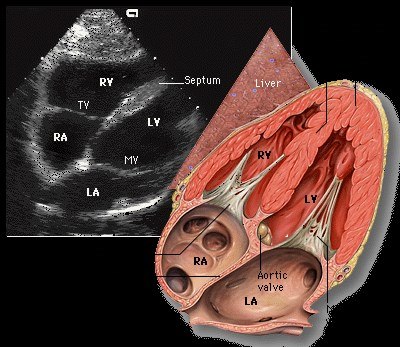
Size is the first and fastest read. The right ventricle normally sits smaller than the left, around two-thirds its width, and the left ventricle alone forms the point of the heart. A glance at the four-chamber view answers whether that holds. In cross-section the healthy right ventricle wraps around the left as a slim crescent. When the right chamber swells to match the left, loses that crescent shape, or pushes past it to take over the apex, it has enlarged under a load it cannot handle.
The ratio puts a number on the glance. The width of the right ventricle against the width of the left, measured across the chamber at end-diastole, runs near two to three under normal conditions. A ratio approaching one means the right chamber has grown to the size of the left. A ratio past one means it has overtaken it, the picture of a right ventricle straining hard. A handheld reader who eyeballs the two chambers side by side reaches the same call the measured ratio gives, fast enough for a crashing patient. The size read alone does not date the trouble, since a long-standing strain and a sudden one can both widen the chamber. That is the job of the squeeze and the wall thickness that follow, so size opens the read and the later findings finish it.
Size opens, the rest dates it
Size flags a strained chamber in a glance. Whether the strain is recent or old waits on the squeeze and the wall that follow.
Does it still squeeze
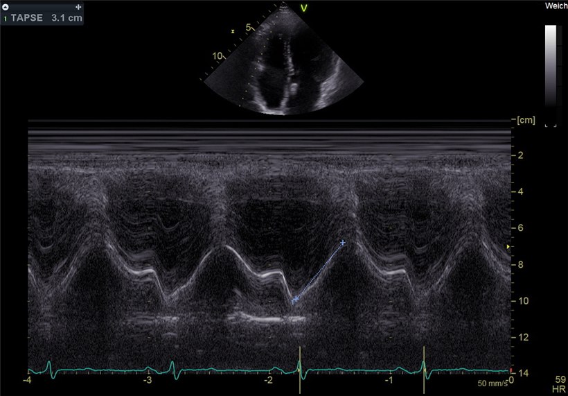
A wide chamber that still pumps is a different problem from a wide chamber gone slack, so the next read is the strength of the squeeze. The right ventricle contracts in good part by pulling its base down toward the apex, a lengthwise wringing. The left ventricle grips inward; the right wrings down its long axis. That lengthwise motion is what the bedside measures. The tool is called TAPSE, the travel of the tricuspid ring toward the apex with each beat. An M-mode line laid through the outer corner of the tricuspid valve draws that travel as a moving trace, and the height of the trace is the excursion in centimeters. A ring that drops well over a centimeter and a half with each beat marks a right ventricle still squeezing. A ring that barely moves marks one that has failed, whatever its size. TAPSE reads one corner of the ring rather than the whole chamber, so it stands as a shorthand for the global squeeze, quick to capture and easy to repeat on a small screen. A reader who lays the M-mode line straight along the direction the ring travels reads it true. A line set across that direction reads the excursion short.
The sign that points at the lungs
An acute clot in the lungs loads the right ventricle in minutes, faster than the chamber can adapt, and that sudden strain leaves a signature a handheld scan can catch. The clearest is the way the strained chamber moves. In acute pulmonary embolism the free wall of the right ventricle goes still as the apex keeps contracting hard, a split pattern named for the physician who described it. The mechanism behind it explains why it points so squarely at the lungs. A clot lodged in the pulmonary arteries throws a sudden high pressure at a chamber built only for low pressure, so the thin free wall, bearing the brunt, gives out within minutes. The apex sits anchored to the hard-working left ventricle next door, which tethers it and keeps it moving even as the free wall fails, so the contraction splits into a dead wall and a lively tip. That split is what a reader looks for. Studies put the pattern at roughly three-quarters sensitivity and well over ninety percent specificity for an acute clot, with a high negative predictive value, so its presence argues hard for pulmonary embolism and its absence does not rule the diagnosis out. A reader who sees a stilled free wall with a beating apex in a patient short of breath has a strong bedside pointer toward the lungs, enough to move the workup forward before any scan returns. The finding carries weight because it is hard to fake. A chronically strained right ventricle, one that has thickened and adapted over months, tends to move poorly all over, without the clean split of a dead wall and a live apex, so the split itself flags the strain as recent. The reader still treats the sign as a strong suspicion, short of a settled diagnosis, since a few other sudden insults to the right heart can mimic the pattern, then confirms it against the rest of the scan plus the clinical picture before acting on it. The handheld probe makes the finding reachable at the bedside, on the patient too unstable to move to a scanner, which is the setting where catching it early changes what happens next. A breathless patient whose right ventricle is wide and split in this way may be a candidate for clot-busting treatment, a decision that hangs on recognising the strain fast, so the seconds the bedside scan saves are the seconds that change the outcome for that patient.
The strained right ventricle also bends the wall it shares with the left. A right chamber under high pressure pushes the septum across, so the normally round left ventricle squashes into the shape of the letter D in cross-section. A D-shaped left ventricle on a short-axis view is the print of a right side pushing harder than the left, the partner finding to the swollen chamber and the stilled wall.
The wall thickness sorts the sudden strain from the long-standing kind. The right ventricle answers a chronic high pressure by thickening its wall over months, so a thick free wall points to a problem the heart has carried for some time. A thin wall on a hugely dilated chamber points instead to a strain that arrived too fast for the muscle to build, the pattern of an acute clot. A handheld reader who notes the wall alongside the size places the trouble in time as well as in space.
Reading the right heart as a set
No single one of these findings carries the weight alone, so a handheld reader reads them as a set that agrees or disagrees. A wide chamber, a low TAPSE, a stilled free wall, a D-shaped septum, all together in a breathless patient build a strong case for acute right heart strain. One finding on its own carries far less, since a wide right ventricle can be an old finding and a single odd beat can fool the eye.
The great vein below the heart completes the set. A right ventricle failing against a high pressure backs that pressure up into the inferior vena cava, so a wide, motionless vein is the expected partner to a strained right chamber. A vein that still collapses normally points away from a right heart under serious acute load, a useful check when the chamber size is borderline. The two readings, taken in the same minute from the same window, lean on each other.
The order of the sweep keeps it fast. A reader takes the four-chamber view first, since size and the stilled-wall pattern both read off it, then drops the M-mode line for TAPSE without moving far, then swings to the short axis for the septum, then tilts down to the vein. Four windows, one continuous motion of the probe, under a minute in trained hands. The economy is what lets the read happen at all on a patient who cannot spare longer.
The right ventricle answers fast or it answers not at all. A reader who gathers those reads in one sweep, the chamber and its squeeze, the septum and wall, the vein behind them, holds a picture of the right heart that a single number could never give, assembled in the time a sick patient allows.
Where the bedside read stops
The handheld read is a screen, not the last word, so it is taken for what it can do. TAPSE drops after heart surgery for reasons that have nothing to do with right heart failure, so a post-operative low reading is read with that caveat. The stilled-wall pattern of an acute clot can be mimicked by other sudden strains on the right heart, so it raises suspicion and stops short of settling the diagnosis. A poor four-chamber view, common on a sick patient who cannot lie flat, throws the size ratio off, since a foreshortened chamber reads the wrong width. The same view exaggerates the right ventricle when the probe cuts it obliquely, so a chamber that looks alarmingly wide on one angle settles to normal on a cleaner cut, which is why a reader hunts for the truest four-chamber plane before calling a chamber enlarged.
The findings also lag behind a slow problem. A right ventricle failing gradually compensates and remodels, so its numbers drift slowly over months. A single bedside look catches only where the chamber sits that day. Trending the size and the TAPSE across visits tells the slow story a single scan cannot.
The handheld view is also coarser than a full study, so it carries the qualitative findings well and the fine numbers less surely. A glance separates a normal right ventricle from a grossly strained one with confidence. A borderline TAPSE, a millimeter either side of the cutoff, asks more of the small screen and the steady hand than a pocket device reliably gives, so a marginal number is read as marginal, never as a verdict.
The screen sees the right heart. It does not see the clot.
Read inside those limits, the right ventricle is one of the highest-yield things a pocket probe checks on a breathless or a shocked patient. Those few quick reads, gathered in a single bedside sweep, turn a vague worry about the right heart into a picture clear enough to act on. That is a strong return for a probe in a pocket and a minute at the bedside.
Common questions about the right ventricle on POCUS
What does a handheld right-ventricle check answer?
Three things in seconds: whether the chamber is enlarged, whether it still squeezes, and whether a clot has loaded the lungs in front of it. None needs a tracing or a calculation.
How do you tell the chamber is enlarged?
The right ventricle normally sits about two-thirds the width of the left and stays off the apex. A ratio approaching one, a lost crescent shape, or a chamber taking over the apex marks enlargement under a load.
What is TAPSE?
The travel of the outer tricuspid ring toward the apex with each beat, drawn by an M-mode line. A ring that drops well over a centimeter and a half marks a right ventricle still squeezing; one that barely moves marks failure.
What does the McConnell sign show?
In an acute pulmonary embolism the free wall goes still while the apex, tethered to the left ventricle, keeps beating. The split has roughly three-quarters sensitivity and over ninety percent specificity for an acute clot.
What is the D-shaped septum?
A right ventricle under high pressure pushes the shared septum across, so the round left ventricle squashes into a D in short-axis view, the print of a right side pushing harder than the left.
How is an acute strain told from a long-standing one?
Wall thickness dates it. A thick free wall points to months of high pressure; a thin wall on a hugely dilated chamber points to a strain that arrived too fast for the muscle to build.

About the Author
Julien Mercier
Senior R&D Engineer · Medical Ultrasound Transducer Development
Senior R&D Engineer with an M.S. in Applied Physics and over 15 years of experience in medical ultrasound transducer development, specializing in the design verification and performance testing of high-frequency imaging transducers. Currently leading the development and verification of the company’s next-generation high-frequency linear-array transducer, responsible for imaging performance evaluation and reliability analysis in preclinical testing. Brings extensive hands-on experience in piezoelectric element tuning, beamforming parameter optimization, and system-level performance testing.