


































Volume status
Inferior Vena Cava IVC Assessment for Volume Status with Handheld Probe
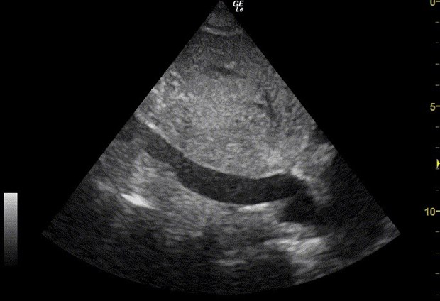
How full the circulation is shows in the width of one vein. The inferior vena cava, returning blood to the right heart, stays soft and narrow when pressure is low and caves in as the chest draws breath; backed up behind a struggling heart, it sits wide and stiff and barely stirs. A handheld probe below the breastbone reads that width and that movement in seconds, turning a question about a patient’s fluid state into two numbers the bedside can act on.
Finding the vein and measuring it right
The probe sits below the breastbone, angled up under the ribs. The reader rocks it until the vein appears as a long dark channel running through the liver toward the right atrium. The measurement is taken a short way back from where the vein meets the heart, about two centimeters downstream, past the point where a liver vein joins. A caliper set too close to the heart catches the funnel where the vein flares into the atrium, which reads wider than the vessel itself.
One check comes before any measurement: telling the vein from the aorta that runs alongside it. The aorta is round and thick-walled, pulsing hard with the heartbeat. The vena cava is thinner-walled. It changes shape with the breath and drains the liver’s own veins into its side. A reader who measures the artery by mistake reports a number that means nothing about volume, so the identity of the tube is settled before the caliper goes near it.
One mistake accounts for the bulk of bad readings. A plane that slices across the tube at a slant catches a short oval, never the full width, so the diameter reads small. The cure is to swing the probe until the vein runs its whole length across the screen with both walls parallel, then measure straight across at the right spot. A vein that tapers or fades at one end of the screen is a plane sliding off the vessel, not a true picture of its width. A reader who confirms the long axis first spends nothing extra and removes the commonest source of a wrong number.
Reading the width against the collapse
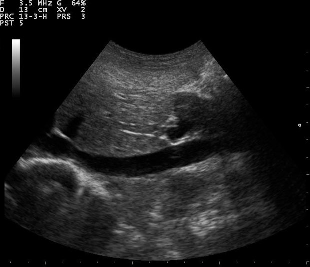
Two numbers come off the vein, and they answer together. The first is the widest diameter, taken at the end of a quiet breath out, when the vein is fullest. The second is how far the vein narrows when the patient breathes in, written as a percentage of that widest width, the figure clinicians call the collapsibility. A patient breathing normally drops the pressure inside the chest with each breath in. That drop sucks a soft low-pressure vein sharply inward, so a big collapse marks a low filling pressure. A vein held open by high back pressure scarcely moves. The two readings sort into a rough map of the pressure on the right side of the heart. A vein under about two centimeters wide that collapses more than half its width points to a low filling pressure, near the bottom of the normal range, the picture of a circulation with room for fluid. A vein over about two centimeters that barely moves points to a high filling pressure, the picture of a circulation already backed up against a heart that cannot clear it. The broad middle, a vein near two centimeters with a moderate collapse, covers the majority of patients, which is why the reading is reported as a band, not a single pressure. A sniff, one sharp inward breath, deepens the collapse and helps when a quiet breath leaves the answer flat. The whole reading rests on a single physiological fact: the soft-walled vein transmits the pressure just upstream of the heart, so its size and its give report that pressure without a needle. The number it stands in for, the right atrial pressure, once needed a catheter threaded into a central vein to read directly. A handheld probe below the ribs now estimates the same pressure from the width and the collapse, the bedside reading a clinician treats as a gauge of whether the tank runs empty or overfull. The measurement is anchored to the breathing cycle, so the widest diameter is read at the end of a quiet breath out and the narrowest at the depth of a breath in, the two extremes the cycle offers. An M-mode line dropped through the vein draws those two widths as a single moving trace, the same trick that times the heart’s walls, which makes the collapse easy to read off one frozen image. The reader no longer chases it between live frames. A reader who lacks a steady breath-hold from the patient takes several cycles and reads the clearest one.
The estimate is a guide, not a gauge calibrated to the millimeter. A reader reports it in the language of low, indeterminate, or high filling pressure, since the bands are what a fluid decision turns on. A precise-looking pressure carried to a single figure claims more than the soft vein can support.
Why the reading turns over on a ventilator
Everything so far assumes a patient breathing on their own. A ventilator inverts the physics. A machine that pushes air into the lungs raises the pressure inside the chest with each delivered breath, so the vein widens on the breath in where a spontaneous breath would have collapsed it. The number a reader watches becomes the distension, how far the vein swells as the ventilator inflates, the mirror image of the collapse a breathing patient shows. That inversion is a trap for a reader who forgets it. The same vein, the same probe, the same swing of diameter on the screen can mean opposite things depending on who is driving the breath. A reader checks how the patient is breathing before naming what the movement means. The shared thread across both states is the size of the swing. A vein that moves a lot with breathing, collapsing in a spontaneous patient or distending under a ventilator, tends to mark a circulation that will respond to a fluid bolus. A vein that holds nearly still tends to mark one that will not, since its pressure no longer rides up and down with the breath. The respiratory swing is where the static vein touches the dynamic question that a stroke-volume test answers from the other side. The two readings, the vein’s movement and the change in forward flow, look at the same fluid question through different windows, and a reader who has both trusts a conclusion they agree on more than either alone.
Read the breathing before the swing
Which way the breath drives the vein decides what its movement means. A reader settles that before reading the swing.
The bedside questions the vein answers fastest
The vein does its fastest work on a short list of recurring questions. The first is the undifferentiated low blood pressure. A flat, collapsing vein steers that patient toward fluid plus a search for bleeding or dehydration. A full, still vein steers the same low pressure away from fluid and toward a pump or an obstruction, a heart that cannot move what it already holds. The vein sorts those two paths in seconds, before any slower test returns.
The second is the breathless patient on a dialysis or a heart-failure background. A wide, motionless vein backs up the clinical impression of fluid overload with a number. It also gives a target to watch as fluid is drawn off, the vein narrowing and starting to move again as the load comes down. A dialysis unit reads the vein before and after a session to judge how much to remove.
The third is the patient already receiving fluid, where the question is when to stop. A vein that was collapsing and has filled out, losing its swing, marks a circulation that has taken what it needs. Pushing past that point only backs pressure into the lungs. The vein gives the resuscitation a brake as well as an accelerator.
None of these is a diagnosis on its own. Each is a fast nudge toward or away from fluid, delivered in the seconds the question is live, by a probe that needs no line and no laboratory. That speed at the decision point is the whole value.
The vein answers fast. It does not answer everything.
What the vein cannot tell on its own
The vein speaks for the right side of the heart, which is not always the side that matters. A left heart failing behind a healthy right one floods the lungs while the inferior vena cava still looks unremarkable, so a normal vein is no promise the circulation is well. The reading answers the right-sided question it was built for and stays silent on the left.
Several everyday things throw the simple rule off. A young athlete carries a wide vein on a low pressure, the build of a trained venous system mistaken for overload. A patient straining, coughing, or breathing hard swings the vein for reasons unrelated to volume. A tense or distended abdomen presses on the vein and fakes a narrowing. Each of these breaks the clean link between size and pressure, so the number is checked against how the patient looks and what the rest of the scan shows before it is believed.
The reading also drifts with the probe. An angled plane shrinks the apparent width, a gate measured at the wrong level catches the wrong calibre, a heavy hand on the belly can squeeze the vein the reading is meant to observe. These are the same handling errors any vein measurement carries, and they are why a careful reader repeats the view before committing to a number that will steer a liter of fluid in or hold it back.
Held inside those limits, the inferior vena cava is one of the quickest high-value reads a pocket probe offers. It places a sick patient on the scale from empty to overloaded in seconds, points toward or away from fluid, and stands in for a measurement that once cost an invasive line. Measured on its long axis, at the right spot, the width read against the collapse, the breathing accounted for, the answer taken as a band against the whole patient, it pulls a real clinical signal from a single dark stripe under the liver. That is a great deal of information for a probe in a pocket and a few seconds at the bedside.
Common questions about the IVC assessment
What does the inferior vena cava tell you?
Its width and how far it moves with breathing stand in for the filling pressure on the right side of the heart, a window onto how full the circulation is, read without a central line.
Where is the vein measured, and how is it told from the aorta?
About two centimeters downstream of the heart, past where a liver vein joins, on the long axis. The aorta is round, thick-walled and pulsing; the vena cava is thinner-walled and changes shape with the breath.
How do the width and collapse read volume?
A vein under two centimeters that collapses more than half points to a low filling pressure with room for fluid. A vein over two centimeters that barely moves points to a high pressure already backed up. The reading is taken as a band.
Why does a ventilator reverse the reading?
A ventilator raises the chest pressure on the breath in, so the vein distends where a spontaneous breath would collapse it. The reader watches the distension instead of the collapse and checks how the patient breathes first.
What throws the reading off?
A trained athlete carries a wide vein on a low pressure, a tense abdomen fakes a narrowing, and straining or coughing swings the vein for reasons unrelated to volume. The number is checked against the whole patient.
Does a normal vein mean the circulation is well?
Not on its own. The vein speaks for the right side of the heart; a left heart failing behind a healthy right one can flood the lungs while the vein still looks unremarkable.

About the Author
Julien Mercier
Senior R&D Engineer · Medical Ultrasound Transducer Development
Senior R&D Engineer with an M.S. in Applied Physics and over 15 years of experience in medical ultrasound transducer development, specializing in the design verification and performance testing of high-frequency imaging transducers. Currently leading the development and verification of the company’s next-generation high-frequency linear-array transducer, responsible for imaging performance evaluation and reliability analysis in preclinical testing. Brings extensive hands-on experience in piezoelectric element tuning, beamforming parameter optimization, and system-level performance testing.