


































Cardiac function
Left Ventricular Internal Dimension LVID Handheld Ultrasound Measurement
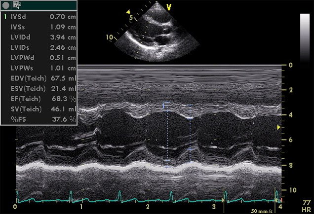
One caliper line drawn straight across the main pumping chamber, septum to back wall, is the oldest measurement in cardiac ultrasound and still one of the steadiest. That line is the left ventricular internal dimension, read twice: at the chamber’s widest in diastole, written LVIDd, and its narrowest in systole, written LVIDs. From those two numbers a handheld scan reads whether the ventricle is enlarged, whether its walls are thick, and how hard it squeezes. On a small probe the single line stays steady where traced volumes often cannot.
Where the caliper line belongs
The measurement starts in the parasternal long-axis view, the probe between the ribs at the left sternal edge, the ventricle laid out lengthways on the screen. The caliper crosses the chamber at right angles to its long walls, placed at the level where the mitral valve leaflet tips reach into the cavity. That landmark matters. Measure too high toward the valve ring and the chamber reads wider than it is. Measure too low toward the apex and it reads narrow. The mitral tip level is the agreed spot that lets one reader’s number compare with another’s.
The level also carries a quiet revision behind it. The traditional measurement sat lower, at the level of the chordae tendineae below the valve, the spot a blind M-mode line tended to find. Current guidance moved the landmark up to the mitral tips and asked for the line to be placed on a two-dimensional image, the shift that lets the number from a modern handheld scan line up with the reference ranges a clinic quotes.
Perpendicular is the other half of the rule. A caliper line that crosses the chamber at a slant cuts a longer path than the true width, so a tilted measurement reads too large every time. The ventricle has to lie flat and lengthways before the line goes down, so the view is worked until the chamber sits horizontal on the screen with both walls clean. A handheld probe makes that alignment the whole task, since a rushed oblique view feeds an inflated number into every calculation that follows. A line dropped square to the walls is the difference between a true width and one read several millimeters long.
Timing within the beat is the last placement rule. The diastolic width belongs to the frame at the foot of the QRS complex on the trace, the instant the chamber is fullest before it contracts. The systolic width belongs to the frame where the back wall has climbed closest to the septum, the bottom of the squeeze. A reader who keeps the electrocardiogram trace running at the foot of the image has a timing reference for both, since the eye alone can misjudge the fullest and emptiest frames by enough to shift the shortening figure a few points either way.
M-mode against a frozen frame
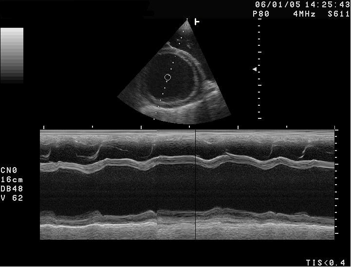
There are two ways to lay the caliper down. The old way is M-mode, a single scan line sampled hundreds of times a second and drawn as a moving graph of depth against time. The walls of the ventricle show up as bright bands that march toward each other in systole and apart in diastole, so a reader picks the widest and narrowest points off one clean tracing. M-mode samples so fast that the exact moment of fullest and smallest chamber is caught with a sharpness a frame-by-frame image cannot match, which is why the trace still holds its ground for a measurement that lives on catching those two instants.
The newer way is a direct caliper on a frozen two-dimensional frame. Modern guidance leans toward this. The M-mode line is fixed straight down the screen, the ventricle rarely lies in that exact direction, so the old trace often crosses the chamber at a slight slant and reads high. Dropping calipers on a paused two-dimensional image lets the reader place the line square to the walls, at the cost of the timing sharpness the M-mode trace gives for free. A careful handheld reader uses each for what it does well: the frozen frame for the cleanest square line, the M-mode for the surest grip on the systole and diastole instants. Neither is automatic on a small screen, so the reading is only as honest as the moment the reader stops the loop on.
What the two numbers tell once they are caught
A widened diastolic dimension is the headline a quick scan looks for. A normal adult left ventricle measures roughly four to five and a half centimeters across in diastole, a touch larger in men than in women, so a chamber reading well past that has dilated, the slow remodelling a weakened or volume-loaded heart goes through. A tall patient carries a larger heart, so the raw centimeters are often divided by body surface area before a borderline case is called, the step that keeps a big frame from being labelled diseased. A handheld probe at the bedside flags a dilated ventricle in seconds, the finding that turns a vague breathlessness into a cardiac referral. The two dimensions together also give a contraction index with no tracing at all. Fractional shortening is the diastolic width minus the systolic width over the diastolic width, the share of its own diameter the chamber surrenders with each beat. A heart that goes from 3.9 centimeters to 2.5 shortens by a bit over a third, inside the healthy band of roughly a quarter to a touch above four in ten. A figure below that band marks a weak squeeze. A figure above it marks a hyperdynamic heart. Fractional shortening reaches an answer fast and holds steady on a small screen, since it rides on two caliper points where the traced figure rides on a hand-drawn outline. The speed brings a temptation, the Teichholz calculation, an old formula that projects a whole-chamber volume and an ejection fraction from the single diastolic and systolic width, the figures that still print on the M-mode readout. The projection assumes the ventricle is a tidy geometric solid that scales evenly from one measured width. A healthy heart roughly obeys that assumption. A diseased one breaks it, so the printed volumes hold only when the chamber is symmetric. The limit is built into how few points the scheme rests on. Fractional shortening reads only the width at one level near the base of the heart. A ventricle that fails in one wall while another wall still moves can post a normal figure that flatters it: a heart attack that kills the far tip leaves the basal width swinging almost normally, so the shortening figure misses the dead apex. This is the price of the single line, fast and steady at one level, then quiet about trouble that sits away from it. A reader who knows that limit treats a normal fractional shortening in a sick patient as a reason to look wider.
What one line cannot see
The single dimension reads fast and steady at one level. A wall that fails away from that level can slip past it unseen.
Reading the walls in the same line
The caliper line that catches the chamber also crosses the muscle on each side, so the same measurement reads the septum in front and the back wall behind. A wall past roughly a centimeter at end-diastole points to hypertrophy, the muscle bulked up against years of high pressure or a thickening disease. A handheld scan that reads a thick wall alongside a small tight cavity sees the pattern of a pressure-loaded heart in one frame. A thin wall stretched around a wide cavity is the opposite pattern, the dilated failing heart. The dimension line, read across all four structures it crosses, sketches the shape of the disease before any other measurement is taken.
The wall and the cavity together carry one more index a reader builds in the head. The ratio of wall thickness to chamber radius separates two kinds of thickened heart: a heart that has grown its walls inward around a normal or small cavity, the concentric pattern of long-standing pressure, from a heart that has thickened while its cavity stretched, the eccentric pattern of volume overload. A handheld reader who notes both the wall and the width reads that pattern off the same caliper line, a piece of diagnosis the traced volume never offers, since the traced figure reports the cavity alone and stays quiet on the muscle around it. The wall reading has its own trap to dodge. The caliper has to start at the true inner edge of the septum and stop at the true inner edge of the back wall, so a line that catches a fold of the right ventricle in front, or a smear of pericardial fat behind, reads a wall thicker than the muscle on its own. Reading the thickness at end-diastole on a clean line, with the chamber at its widest, keeps the wall figure honest the same way it keeps the cavity figure honest. One disciplined line serves both.
The wall reading carries the same perpendicular rule as the cavity. A slanted line thickens the apparent wall the way it widens the apparent chamber, so the squareness worked out for the cavity measurement serves the wall measurement at no extra cost. One clean line, dropped square at the mitral tip level on a flat long-axis view, hands a handheld reader the chamber width, the contraction, and the wall thickness together. That economy is why the oldest measurement in the field still opens the bedside study of the left ventricle, ahead of the traced volumes that need a clearer image and a steadier hand than a pocket probe often allows.
Why the old line outlasts the new one
The linear measurement predates the traced volume by decades, and the newer method was supposed to retire it.
It did not, for a reason that suits the handheld probe exactly. The traced volume needs a clean endocardial border all the way around the chamber, a border a small noisy screen often cannot deliver. The caliper line needs only two clear points, the septum and the back wall at one level, points that survive a rougher image. A scan that could never support a reliable traced outline can still drop an honest dimension line, so the old method reaches a usable number on hardware that defeats the new one.
The number also trends well across visits, which matters for the chronic heart a clinic follows. A dilated cavity that grows two millimeters between scans is a real signal. The caliper catches that drift more repeatably than a hand-traced area whose outline shifts with whoever drew it. A reader following a failing heart over months leans on the dimension for that steadiness.
Its honest place is as the opener, not the whole study. The dimension line flags the dilated chamber, the thick wall, the weak shortening in the seconds a bedside scan can spare, then hands off to a fuller assessment when the finding warrants it. A handheld probe used this way spends its limited image quality where that quality goes furthest, on two sharp points instead of a long fragile outline.
The reading a pocket scan can defend is the one its picture can support. For the left ventricle, more often than the textbooks built around cart machines admit, that reading is a single square caliper line at the mitral tips, read for all three things it crosses.
Common questions about the LVID measurement
What is the left ventricular internal dimension?
It is the width of the main pumping chamber, measured straight across from the septum to the back wall, read at its widest in diastole (LVIDd) and its narrowest in systole (LVIDs).
Where on the chamber is the caliper line placed?
In the parasternal long-axis view, square across the chamber at the level where the mitral valve leaflet tips reach into the cavity. A slanted or off-level line reads the width too large.
M-mode or a 2D caliper?
M-mode samples fast and catches the fullest and smallest frames sharply. A caliper on a frozen 2D frame lets the line sit square to the walls. A careful reader uses each for what it does well.
What is fractional shortening?
The diastolic width minus the systolic width, over the diastolic width: the share of its own diameter the chamber gives up each beat. The healthy band runs from about a quarter to a little above four in ten.
What is a normal chamber width?
A normal adult left ventricle measures roughly four to five and a half centimeters across in diastole, a touch larger in men. A reading well past that points to a dilated chamber.
Why does the single line suit a handheld probe?
It needs only two clear points, the septum and the back wall, which survive a rough image. A traced volume needs a clean border all the way round, which a small noisy screen often cannot deliver.

About the Author
Julien Mercier
Senior R&D Engineer · Medical Ultrasound Transducer Development
Senior R&D Engineer with an M.S. in Applied Physics and over 15 years of experience in medical ultrasound transducer development, specializing in the design verification and performance testing of high-frequency imaging transducers. Currently leading the development and verification of the company’s next-generation high-frequency linear-array transducer, responsible for imaging performance evaluation and reliability analysis in preclinical testing. Brings extensive hands-on experience in piezoelectric element tuning, beamforming parameter optimization, and system-level performance testing.