Abdominal Ultrasound Scan Sequence From Right Upper to Left Lower Quadrant
A belly scan that finds everything runs the same road every time, beginning under the right ribs and ending in the pelvis. The route is not idle habit: it starts where the windows are easiest and the organs richest, crosses the midline to the great vessel and the buried pancreas, climbs to the awkward high corner of the spleen, then drops to the bladder and the low spaces where fluid settles. Each stop is a station with its own target, its own patient position, its own held breath. A clinician who learns the road as a fixed sequence carries the checklist in the hand, not the head, and a belly read in that order gives up few of its secrets to the probe.
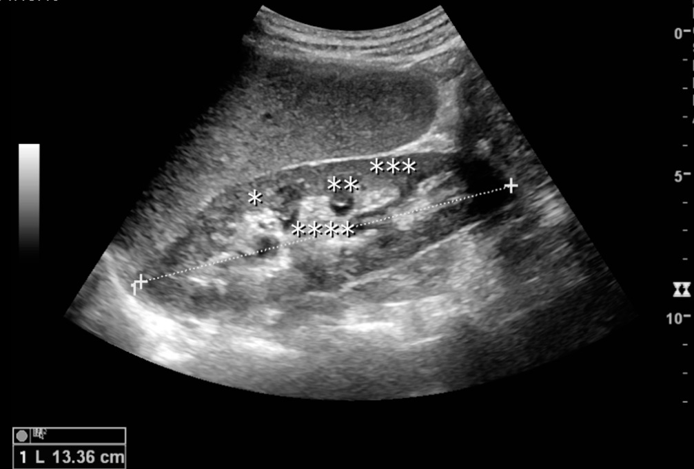
A normal kidney in long axis: the flank station, scanned just after the liver on the way down the road. Credit: Wikimedia Commons (CC).
One road, the same way every time.
From the right ribs to the pelvic floor: four stations walked the same way every time.
How the curved probe makes its picture, the grey ladder that tells liver from kidney, the tricks that beat the gas: these sit in the overview on the convex abdominal exam. The road itself is the work here, station by station from the right ribs to the pelvic floor.
The right upper quadrant: the first station
The journey opens under the right costal margin, the busiest stop on the whole road and the one a hurried scan shortchanges first. The liver comes up first, the probe laid subcostal or slipped between the ribs and fanned through the right lobe, the smooth edge, the dome tucked under the diaphragm, the great vein running in toward the heart. The gallbladder follows, found by sliding down and a little medial and turning the probe along its long axis then across it, often with the patient rolled onto the left side or holding a deep breath to drop it clear of the colon that loves to sit in front of it. The right kidney sits just behind and below, swept along its length and turned through its short axis, its bright centre and darker rim read against the liver beside it. Last comes the hepatorenal recess, the dip between liver and kidney that is the lowest point of the upper belly in a patient lying flat, where a first thin black stripe of free fluid shows; that check takes seconds and its trauma reading lives in the pages on the focused fluid scan. The order inside the station carries its own logic: liver first to set the grey reference the rest are judged against, gallbladder steadied by the held breath, kidney and recess last as the probe slides down off the liver. Two or three positions and a couple of breath-holds are spent here before the road moves on, since this one corner answers a large share of the questions the whole scan is asked.
The discipline of the station is to finish it before leaving. A probe that wanders off to the midline with the gallbladder half-seen leaves a gap the rest of the road will not fill, and the right upper quadrant, richer than any other stop, punishes a half-look harder than any. Liver, gallbladder, kidney, recess: four targets, one corner, no leaving until each has shown itself. The temptation to chase a striking finding straight off to another quadrant is the commonest way the first station ends half-done and a quiet target slips by unread.
The right ribs hide the busiest corner of the belly.
The midline: the aorta and the buried pancreas
From the right ribs the probe slides to the centre of the upper belly, where the body’s largest vessel runs the midline ahead of the spine. The aorta is found just left of centre and followed in long axis from below the diaphragm down to where it forks near the navel, then turned across into short axis so its calibre can be read as a clean circle at intervals along its length; that width is the reason the midline is never skipped, and its aneurysm reading is carried in the pages on the aortic screen. The pancreas drapes across the vessels behind the stomach, shy behind a curtain of gas that a swallow of water or a patient press can sometimes lift, the station’s reward when it shows and its frustration when it hides.
The vessels are the map of the midline. The aorta lies ahead and a little to the left, thick-walled and pulsing hard; the inferior vena cava runs to its right, thinner-walled and easily squashed under a press, its calibre rising and falling with the breath. Tell the two apart once at the start, and the pancreas, the vessels’ tributaries, the spine behind all fall into their places around them. Lose track of which tube is which, though, and the midline turns into a puzzle of look-alike vessels that no careful measuring can untangle.
The midline is read around its two great vessels.
The left upper quadrant: the spleen’s awkward corner
The left upper quadrant is the road’s hardest stretch, and the place a sequence is likeliest to fail. The spleen sits high and far back, tucked under the lower ribs and the edge of the lung, reached only by sliding the probe up onto the posterior axillary line, higher and more toward the back than the right-sided window ever needed. A deep breath drops it a little, a roll onto the right side brings it down further into reach. The left kidney hides behind and below the spleen; the splenorenal recess between them is the left twin of the hepatorenal dip, another low pocket where fluid gathers and another quick check on the way past.
Left is higher than the eye expects, and that single fact rescues a failing left-upper view. A probe set where the right-side window sat finds only rib and lung and a frustrated operator; slid up two interspaces and angled back toward the spine, the same probe brings the spleen into the frame. The window stays narrow and the rib shadows many, so the station rewards a slow hand and a patient breath more than any other.
The spleen sits higher and further back than the hand expects.
Down to the pelvis
The road ends low, in the pelvis, where the bladder makes its own window. Read full, the bladder is a black box of urine that throws a clear path down onto the space behind it; emptied, it collapses and the pelvis turns murky. Behind the bladder lies the lowest point of the whole abdomen in a patient lying flat, the rectovesical pouch in a man and the pouch of Douglas in a woman, the final sink where free fluid pools after everywhere higher has drained into it. A scan that has run from the right ribs to this pocket has swept the belly’s high ground and its low drain both.
The pelvis is the belly’s drain, and the reason the road ends there rather than at the spleen. Whatever leaks or bleeds or weeps higher up runs downhill to the lowest space the body offers, and lying flat that space is the pocket behind the bladder; a clean pelvis at the close of the sweep is a reassurance the upper stations on their own cannot give, the dependent recesses checked in the order fluid would have filled them.
The pelvis is where the belly drains.
Holding the thread between stations
The road is only as good as the moves between its stops. The probe is kept sliding on the skin and never lifted clear; the picture then flows from one station to the next and the eye never loses its place; the orientation marker is left untouched through the whole sweep; left and right and near and far keep the same meaning from the first stop to the last. A breath is called at each rib-bound station and let go between them, the lungs working with the probe instead of against it. The transitions are where a careless scan drops a station without noticing, the probe skating past the spleen or the recess as the operator believes the road already complete.
The picture should slide, never jump.
Adapting the road to the patient
The road bends to the patient in front of it. A large abdomen forces the probe to a lower frequency and more positions at every stop, each window harder won than the last; a patient who cannot lie flat reshapes the dependent recesses, since the lowest point of a propped-up belly is no longer the pocket behind the bladder. Fluid hunted in the wrong corner is fluid missed. A pregnant patient brings a crowded pelvis and a uterus that shoulders the route aside, the bladder window read with a gentle hand and the deep press eased right off. A restless child takes the whole road at speed, a higher-frequency window and a quick clean pass before patience runs out.
The order survives even when the positions do not. Whatever the body allows, the sequence keeps its logic: the easy windows first, the great vessel down the centre, the dependent drains last in the order gravity fills them; a road bent to the patient is still a road, checked off from end to end, every station accounted for whatever shape the body takes.
The road bends to the patient; it does not break.
When the order is broken on purpose
In the crashing patient the road sometimes runs out of sequence. A collapse with a rigid belly sends the probe straight to the station likeliest to hold the answer, the aorta in an older patient gripped by back pain, the pelvis and the recesses in one bleeding after a fall, the urgent question answered before anything else. The full sweep follows only once that answer is in hand and the patient is steadier. The order is a default the hand returns to, picked up again the moment the emergency loosens its grip; a sequence known cold is what lets a clinician break it safely and find the place again afterward.
The urgent station first; the rest of the road right after.
Where the sequence misleads
The danger of a fixed road is the belief that finishing it means seeing everything. A station run through a screen of gas, a spleen glimpsed for a single second, a recess searched in a body too deep for the beam: each can be ticked off as done while showing nothing that can be trusted, the sequence complete on paper and the organ unread in truth. The route guards against skipping; it cannot promise sight.
The cure is honesty at each stop, a poor window named as poor and revisited or handed on rather than counted as clear. A sequence walked end to end with three bad windows is three questions unanswered, no matter how tidy the checklist looks; the road is a scaffold for thoroughness, never a substitute for the judgement that knows when a station has truly shown its organ and when it has only pretended to.
Finishing the road is not seeing the belly.
The whole road in one sweep
From the right ribs to the pelvic floor, the abdominal sequence is a single disciplined sweep through a belly full of overlapping organs: the busy right upper quadrant with its liver and gallbladder and kidney, the midline vessel and its shy pancreas, the high hidden spleen and its recess, the low pelvic drain behind the bladder. Run the same way every time, the road turns a crowded, confusing abdomen into an ordered list checked off in minutes, each station handing the probe to the next. It is the scaffold the focused abdominal scan is built on, the plain habit that keeps a fast look from sliding into a careless one and sends the patient onward with the belly’s high ground and its low drain both accounted for. A road this plain is easy to teach and hard to forget, which is why the same sweep, learned once, serves the novice and the seasoned hand alike at the bedside.
About the Author
Julien Mercier
Senior R&D Engineer · Medical Ultrasound Transducer Development
Senior R&D Engineer with an M.S. in Applied Physics and over 15 years of experience in medical ultrasound transducer development, specializing in the design verification and performance testing of high-frequency imaging transducers. Currently leading the development and verification of the company’s next-generation high-frequency linear-array transducer, responsible for imaging performance evaluation and reliability analysis in preclinical testing. Brings extensive hands-on experience in piezoelectric element tuning, beamforming parameter optimization, and system-level performance testing.