Forklift Batteries
Forklift Solutions
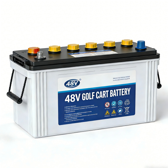
Golf Cart Batteries
Golf Cart Solutions
AGV & AMR Batteries
AGV Solutions
Pallet Jack Batteries
Pallet Jack Solutions