





































Infective Endocarditis Ultrasound Diagnosis Handheld Echocardiography
Infective endocarditis is an infection that settles on a heart valve and builds a clump of germs and clot called a vegetation. A handheld scan looks for that vegetation and the damage it does to the valve. The diagnosis rests on blood cultures and the clinical picture together with the scan. A clinician reads the valve for a mobile growth, a new leak, and the wreckage the infection leaves. A focused scan at the bedside brings the first look at a valve under attack.
What the scan looks for
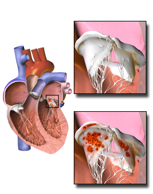
The infection takes hold on a heart valve, often one already damaged or built from man-made parts. Germs settle on the roughened surface and draw in layers of clot, piling up into a soft mass called a vegetation. The vegetation rides the valve through every beat, sheds fragments into the bloodstream, and eats slowly into the leaflet beneath it. A clinician reads the valve for that growth and for the harm it leaves behind. The scan does not name the germ, since blood cultures do that, so the diagnosis rests on the cultures and the clinical story together with the picture. A handheld brings the picture to the bedside in the hours the cultures take to grow. A clinician asks who is at risk, since a worn valve, a replaced one, a past infection, or a needle habit each opens the door to the germs. A clinician reads the valve that already has a problem as the one the germ finds easiest to settle on.
The scan sits at the centre of the diagnosis alongside the blood cultures. A vegetation found on the valve is one of the major signs the diagnosis is built on. The scan also measures the harm the infection has done, the leak it has opened and the tissue it has eaten. A clinician reads the picture to weigh how far the infection has gone and whether the valve can hold. The find at the bedside often turns a fever of unknown source into a heart infection a clinician can name. The scan carries weight in the diagnosis few other bedside tests can match. A clinician reads it as the one bedside test that can see the infection at work on the valve.
The handheld reads the valve from the chest wall, down to a floor set by the small and the hidden. A small growth, an early pocket of pus, and an infection on a man-made valve all sit below that floor, each one waiting for the deeper study. A clean handheld study in a patient with a strong story never clears the infection on its own. A clinician reads a normal bedside scan as one step on the way to the deeper look. The chest scan catches roughly seven in ten native-valve infections. A clinician records the clean scan with the date and the windows, ready for the next look. A clinician keeps the search open after a clean scan, since the growth it missed is the one that still matters.
A clinician sweeps the valves at the bedside of a patient burning with fever. The focused study runs alongside the blood drawn for culture and the search for a source. A vegetation caught early moves the patient straight onto the path for a heart infection. A clean sweep with a strong clinical story sends the patient on for the study down the gullet. A bedside scan sets the first direction in the hour the fever is worked up. A clinician treats a fever with germs in the blood and no clear source as a heart infection until the valves come back clean. A clinician reads a quiet valve in a feverish patient as a reason to look again, since the growth can be small at first.
The vegetation on the valve
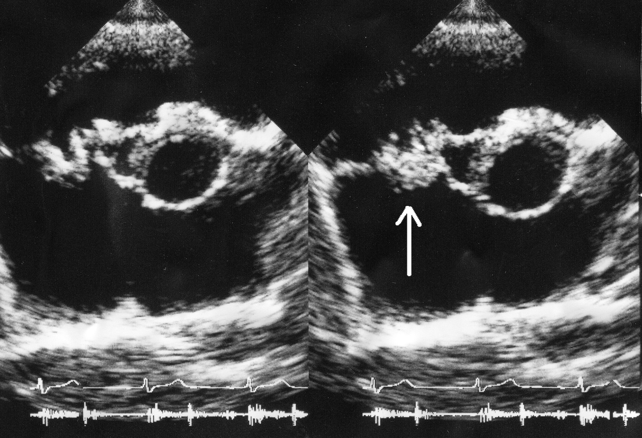
A vegetation shows on the scan as a shaggy mass clinging to a valve leaflet. It carries the brightness of heart muscle, a shade paler than the blood pool around it. The mass keeps to the leaflet itself. Its loose part also swings on its own. A clinician reads that lashing motion, free of the leaflet, as the mark of a vegetation. The growth sits on the surface the blood strikes as it leaks back through the valve. A clinician reads the way the mass moves out of step with the leaflet as the surest sign it is a vegetation.
A vegetation favours the upstream side of the valve, the side the backward jet washes over. On the mitral valve it sits on the atrial face, where a leak drives blood back toward the lungs. On the aortic valve it sits on the ventricular face, where a leak floods back into the pumping chamber. A clinician hunts the vegetation on that low-pressure side, in the path the regurgitant jet carves. The mass swings across the valve when it opens, dipping into one chamber and back. A clinician times that prolapsing motion to the beat, watching the growth ride past the leaflet edge. The shaggy outline and the loose swing set a vegetation apart in the moving picture. A clinician reads the size, the shape, and the wildness of the motion as one description of the growth. The full read takes a slow sweep through the valve, plane by plane, since a vegetation hides in any single still view. A clinician notes that a vegetation on the right side, on the tricuspid valve, turns up often in a patient who injects drugs, throwing its fragments to the lungs. A clinician reads the loose tip of a large vegetation as the part likeliest to tear away.
A clinician separates a vegetation from the growths that mimic it. A lump of calcium sits still, locked onto a thickened leaflet. A torn leaflet flails with a clean edge, free of any clinging mass. A small valve tumour rides a stalk in a heart with no fever and no germ in the blood. A clinician reads a shaggy mass that swings loose, in a feverish patient with germs in the blood, as the one that points to infection. The clinical story carries the read as much as the shape on the screen. A clinician leans on the fever and the germ in the blood to tell a true vegetation from an old, sterile lump. A clinician reads a thrombus or a strand of old tissue as a quiet mass in a heart with no fever and no germ in the blood.
A clinician measures the vegetation across its longest span. A growth past about ten millimetres carries a real risk of shedding a fragment into the circulation. Past about fifteen millimetres, a growth that swings loose carries a risk higher still. A clinician reads the size and the mobility together as a gauge of how likely the growth is to break off. The number feeds straight into the talk about whether the valve needs surgery. A clinician reads the embolic risk as highest in the first days, before the antibiotics have had time to firm the growth. A clinician reads a vegetation that keeps its size on treatment as a reason to stay watchful for an embolus.
A clinician reads the man-made valve and the pacing wire with extra care. A vegetation on a pacemaker lead rides the wire through the right heart, a thin growth on a thin line. A vegetation on a replaced valve hides in the bright glare the metal throws across the picture. A clinician who suspects an infection on a device or a prosthetic valve leans on the study down the gullet. The chest scan rarely clears a man-made valve on its own. A clinician reads a new delay in the heart’s wiring as a hint the infection has burrowed into the aortic root near the conduction path.
The damage the infection does
The vegetation eats into the leaflet it sits on. The infection bores through the thin tissue, opening a hole or tearing a cusp loose from its hinge. The valve now leaks through the gap the infection has carved. A clinician reads the ruined leaflet and the leak it lets through as the structural harm of the disease. A clinician reads the speed of that destruction as the reason the infection turns dangerous so fast.
The new leak shows on colour Doppler as a backward jet of blood through the valve. A clinician lays the colour box over the valve and reads the width and the reach of the backward flow. A heavy leak that opens over days gives the heart no time to stretch and cope, so it floods the lungs and drops the pressure. A clinician reads a severe leak in a feverish patient as a warning the valve is failing fast. A clinician reads the flooded lungs and the dropping pressure as the signs a sudden leak has overwhelmed a heart with no time to adapt. A clinician reads how poorly the heart bears a leak that arrives all at once, since it has not had the months a slow leak allows.
The infection can burrow past the leaflet into the tissue around the valve. A pocket of pus, an abscess, builds in the root of the aorta or the ring of the valve, a swelling beside the leaflet. A clinician reads an abscess as a sign the infection has broken its banks, a finding that calls for surgery. The chest scan catches an abscess late, so a clinician carries the suspicion to the study down the gullet. A clinician reads a pocket near the aortic valve with special care, since the root abscess sits close to the heart’s wiring and the great vessels.
Reading it on a handheld
A clinician opens the valve from more than one window to corner the vegetation. The long-axis and short-axis views from beside the sternum lay out the aortic and mitral leaflets. The apical view swings the valves into a fresh line for the beam. A clinician sweeps each leaflet slowly through its full arc, since a small growth hides in all but one plane. A clinician returns to the window that showed the growth best, marking it for the next study to match.
A clinician sets the depth and the gain to bring out the fine edge of each leaflet. A vegetation moves, so a clinician reads the moving clip, watching the mass swing across the beats. A tight zoom on the valve fills the screen with the leaflet and the growth riding it. A clinician trusts the moving picture to show the lashing part a still frame would miss. A clinician widens the depth to set the valve in its chamber, then narrows back in on the leaflet for the fine detail.
A clinician lays colour over the valve to catch the leak the infection has opened. A new jet of blood through a ruined leaflet stands out against the still tissue around it. A clinician reads the size of that jet as a measure of how far the valve has failed. A new leak in a patient with a fever and germs in the blood pushes the case toward urgent care. A clinician reads a leak that has sprung up since an earlier scan as a strong pointer to a valve under infection.
A clinician writes down the growth in plain measure for the team and the next study. The note carries the size of the vegetation, the leaflet it sits on, the wildness of its motion, the leak it has opened, and any pocket of pus. A clinician dates the study so the next scan reads against a clear mark. The record turns a moment at the bedside into a baseline the whole team can build on. A clinician saves a clip of the swinging growth, the one record that carries its motion to the next reader. A clinician reads the note back at the next visit and lines the old measure against a fresh scan.
When the chest scan is not enough
A clinician reaches past the chest scan when the suspicion outruns what it can show. The chest scan misses a small vegetation, an early abscess, and a growth on a man-made valve, all hidden from a probe on the front of the chest. A probe passed down the gullet sits right behind the valves and reads them with a clarity the chest view cannot reach. A clinician sends a high-suspicion patient straight on to that deeper study when the chest scan comes back clean.
The Duke criteria and the echo’s place
The diagnosis runs on a set of rules that weigh the evidence together. The modified Duke criteria sort the findings into major and minor, and a diagnosis falls out of the combination. A vegetation, an abscess, a new gap under a prosthetic valve, or a new leak each counts as one of the major echo findings. A clinician reads the scan to supply that major piece of the puzzle. A clinician reads the rules as the frame that keeps two clinicians counting the same evidence the same way. A clinician counts a vegetation, an abscess, a loosened prosthesis, or a fresh leak each as that one major echo line.
The echo and the blood cultures each carry one of the two major signs. A diagnosis grows firm where the two land together, a vegetation on the valve and the matching germ in the blood. The scan on its own names neither the germ nor the infection, so a clinician reads it beside the culture bottles every time. The two halves of the evidence settle the diagnosis as a pair. A clinician reads a culture-negative case with care, since earlier antibiotics or a slow-growing germ can empty the blood bottles. A clinician reads the scan and the bottles as two readings of one disease, each catching what the other misses.
A clinician folds the bedside find into the running tally of criteria. A clear vegetation moves the case a long step toward a firm diagnosis. The handheld supplies the echo half in the minutes the patient is first seen. A clinician carries the find to the team as one column of the evidence the diagnosis stands on. A clinician hands the echo column to the team in the first hour, long before the cultures have finished growing.
| Measure | Number | What it means |
|---|---|---|
| Vegetation raising embolic risk | above 10 mm | a larger growth that may break off |
| Higher embolic risk | above 15 mm | a growth watched even closer |
| Chest scan sensitivity, native valve | about 70% | a clean scan does not rule the infection out |
| Esophageal scan sensitivity | about 90% and up | the test for small growths, abscess, and devices |
| Echo findings counting as a major Duke sign | 4 | vegetation, abscess, new dehiscence, new leak |
The complications the scan watches for
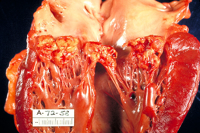
The loose vegetation throws fragments into the circulation, where they lodge and block. A piece carried to the brain causes a stroke, a piece to the spleen or the kidney an infarct, a piece from the right heart a clot in the lung. A clinician reads the size and the swing of the vegetation as the gauge of that danger. The scan watches a large growth that swings free as the one likeliest to break loose. A clinician reads a vegetation that has already thrown a fragment as one that has proven it can do so again. A clinician reads the spleen, the kidneys, and the brain as the places those fragments tend to land.
The infection wears the valve down until the heart begins to fail. A clinician reads the heavy new leak, the spreading abscess, and the prosthetic valve working loose as the marks of an infection out of hand. Each of these turns the case toward surgery, since antibiotics alone cannot rebuild a ruined valve. A clinician carries the structural damage on the scan into the decision to operate. A clinician reads heart failure from a wrecked valve as the leading reason these patients come to surgery. A clinician reads an infection the drugs cannot reach as the case that needs surgery to finish.
Following the infection over time
A clinician scans again through the long course of antibiotics. A vegetation may shrink and firm into a quiet scar once the treatment takes hold. A vegetation that grows on treatment, or a fresh leak, or a new pocket of pus, marks an infection the drugs are losing. A clinician reads each scan against the one before to catch the turn early. The trend across the studies tells a clinician whether the valve is winning or losing. A clinician repeats the study at set points and again at any fresh turn in the fever or the heart. A clinician reads a vegetation that shrinks and firms into a bright scar as the look of an infection settling down.
A clinician brings the scan to the decision on surgery. Heart failure from a wrecked valve, an infection that will not clear, and a large vegetation that has already thrown a clot each push the case toward the operating room. A clinician reads the scan for the evidence the heart team weighs. The picture at the bedside often opens that conversation before the full study confirms it. A clinician reads the scan beside the germ, the fever, and the patient’s strength, since surgery turns on more than one number. A clinician reads the timing as its own question, since an infection caught spreading calls for a faster move to the theatre.
A clinician scans the valve again after the operation or the cure. The repaired valve, or the new one sewn in its place, comes under the beam for its seat and its seal. A clinician reads the function and any leak left behind as the measure of the repair. The post-treatment scan sets the fresh baseline the patient is followed from. A clinician follows a small leak left after a repair across the months after, reading whether it settles or grows. A clinician reads the seat of a new valve and the swing of its leaflets as the measure of a clean repair.
A clinician reads the healed valve as one that carries a longer shadow. A valve that an old infection has scarred sits at a higher risk of a fresh one down the years. A clinician keeps the baseline picture so the next fever has a mark to read against. The record of the valve, kept across the years, makes the next infection easier to catch. A clinician marks the valve a patient was born with or had replaced as the ground every later read stands on.
Infective endocarditis rests its diagnosis on the blood cultures and the clinical story read together with the scan. A handheld brings the echo half to the bedside, finding the vegetation and the damage in the first minutes. A clinician feeds that find into the Duke tally and the decision to operate. The chest scan flags the loud disease. The study down the gullet then settles the quiet case. A clinician reads the valve where the patient lies and carries the hard one forward. A clinician brings the find forward fast, since an infection on a valve costs the patient time it cannot spare.
Common questions about infective endocarditis on a handheld scan
How does infective endocarditis show on an ultrasound scan?
It shows as a vegetation, a shaggy mass that clings to a valve leaflet and swings on it. A clinician finds it on the upstream side of the valve, in the path of the backward leak. The scan also shows the damage, a new leak through a ruined leaflet and sometimes a pocket of pus beside the valve. A clinician reads these together with the blood cultures, since the scan alone does not name the infection.
Can a handheld scan rule out endocarditis?
No. A handheld misses a small growth, an early abscess, and an infection on a man-made valve, all hidden from a probe on the front of the chest. A clean bedside scan in a patient with a strong story never clears the infection. A clinician sends that patient on to the study passed down the gullet, which reads the valves from close behind.
Where on the valve does a vegetation sit?
It favours the upstream, low-pressure side, the side the backward jet washes over. It sits on the atrial face of the mitral valve and the ventricular face of the aortic valve. A clinician hunts the growth on that side, in the path the leak carves. The shaggy shape and the swinging motion mark it out from calcium or a torn leaflet.
What makes a vegetation dangerous?
Its size and its motion. A growth past about ten millimetres carries a real risk of shedding a fragment into the circulation. Past about fifteen millimetres, that risk climbs higher still. A loose, swinging vegetation can break off and lodge in the brain, the spleen, or the kidney. A clinician reads the size and the mobility as the gauge of that danger.
Does the scan diagnose endocarditis on its own?
No. The diagnosis runs on the modified Duke criteria, which weigh the echo findings together with the blood cultures and the clinical picture. A vegetation is one major sign, and a matching germ in the blood is another. A clinician reads the scan as one column of the evidence, carried beside the culture bottles to settle the case.

About the Author
Julien Mercier
Senior R&D Engineer · Medical Ultrasound Transducer Development
Senior R&D Engineer with an M.S. in Applied Physics and over 15 years of experience in medical ultrasound transducer development, specializing in the design verification and performance testing of high-frequency imaging transducers. Currently leading the development and verification of the company’s next-generation high-frequency linear-array transducer, responsible for imaging performance evaluation and reliability analysis in preclinical testing. Brings extensive hands-on experience in piezoelectric element tuning, beamforming parameter optimization, and system-level performance testing.