





































Pulmonary Embolism PE Ultrasound Screening Handheld Echocardiography
A pulmonary embolism is a clot that has travelled to the arteries of the lungs and blocked the blood flow there. A handheld scan reads what that blockage does to the right side of the heart. The clot itself stays out of view, deep in the lung. A clinician reads the strain the clot throws onto the right ventricle and uses it to gauge how dangerous the embolism is. A focused scan at the bedside turns a crashing patient into a picture a clinician can act on in minutes.
What a pulmonary embolism does to the heart
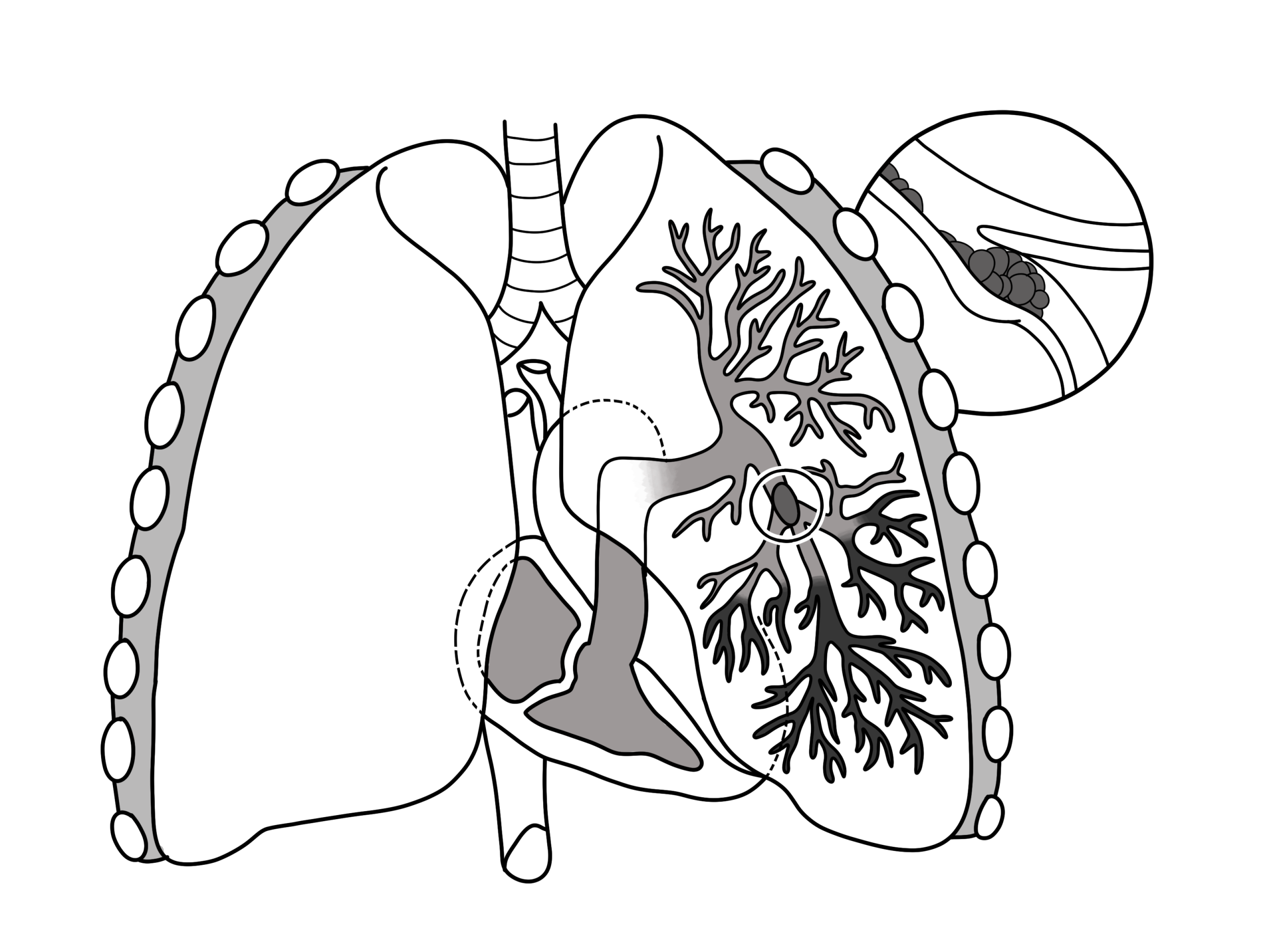
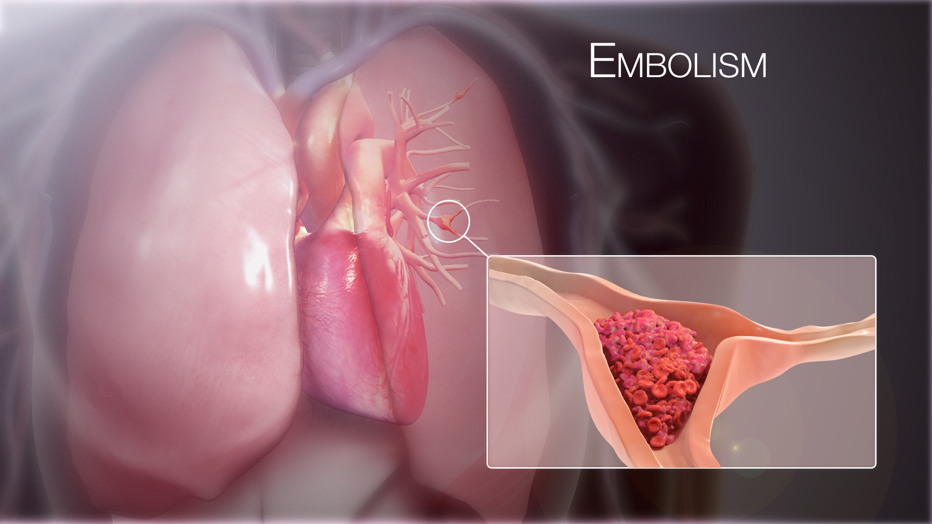
A clot from a deep vein, often in the leg, breaks loose and rides the blood back to the heart. The right side pumps it onward into the lung arteries, where it wedges and stops. Blood can no longer pass that branch to pick up oxygen. The resistance the right ventricle pumps against jumps in an instant. A clinician reads the heart for the strain of that sudden load. The clot has hidden itself deep in the lung, so the scan reads its effect on the right side. A handheld points the beam at the right ventricle and asks one plain question, whether this heart is straining. The body answers the sudden block with a racing heart and a hunger for air, the signs that bring the patient to the door.
The scan earns its place in the patient who is too sick to move. A clinician facing a sudden collapse, a racing heart, and a falling blood pressure needs an answer at the bedside. A picture of a strained right ventricle in that moment supports the call that a clot is behind the crash. The scan arrives in the minutes before any trip to the CT scanner. A clinician can act on a strained right heart in a crashing patient before the lung scan is done. The handheld reads the heart where the patient lies, on the trolley or in the resuscitation bay. A clinician pairs the scan with the story that raised the alarm, the swollen leg, the recent surgery, the long flight in a cramped seat. A clinician reads the scan with the sharpest eye in the patient with cancer, recent surgery, or a long spell in bed, the settings that breed clots.
The scan reads the strain the clot leaves behind. The clot itself sits in the lung arteries, beyond the reach of a probe on the chest. A clinician reads the right ventricle, the chamber that feels the clot’s weight first. A right ventricle that looks normal does not clear the patient of a clot, since a small embolism can settle in the lung and never strain the heart. A clinician treats a normal scan in a suspicious patient as one piece only. The work-up carries on. The strain on the right heart is the shadow the clot casts, the thing the scan can read. A clinician leans harder on the scan where the suspicion runs high and the patient is too unstable for the corridor.
A handheld carries this read into the busiest minutes of a sick patient’s care. A clinician sweeps the right ventricle, the leg veins, and the lungs in one short study at the bedside. The focused look guides the immediate steps. The team arranges the lung scan alongside. A clinician folds the right-heart picture into the decision already taking shape. The full study and the CT confirm the work-up once the patient can travel. A bedside scan moves the first answer to the hour the patient arrives in trouble. A clinician keeps the study short, since a sick patient gives only a narrow window for each view. A clinician learns to read the right heart fast, since a missed clot grows more dangerous the longer it sits.
The right ventricle under sudden strain
The right ventricle does an easy job in a normal heart. It pushes blood the short way into low-pressure lung vessels, so it has a thin wall and a crescent shape that wraps around the left ventricle. A sudden clot load asks it to push against a pressure it has never met. The thin wall cannot thicken in an afternoon, so the chamber answers the only way it can, by stretching. A clinician reads that stretch as the first clear sign of acute strain. The right ventricle swells until it matches or outgrows the left in the four-chamber view from the apex. A normal right ventricle holds about two-thirds the width of the left. A chamber grown to equal the left has already stretched a long way past where it sits in health. A clinician trusts the size read in the four-chamber view, the window that lays the two ventricles side by side for a fair match.
The size of the right ventricle against the left tells the story at a glance. A clinician opens the four-chamber view and compares the two chambers at end-diastole. A right ventricle that reaches the same width as the left has dilated past its normal smaller size. A right ventricle wider than the left has taken on a heavy load. The swelling pushes the wall between the two chambers, the septum, toward the left side. A clinician turns to the short-axis view from beside the sternum, where the left ventricle normally sits as a clean round ring. The overloaded right ventricle flattens that ring along one side and bends it into the shape of the letter D. The flattened septum is the print the strain leaves on the left chamber, plain on a single frozen frame. A clinician reads the D-shape together with the dilated right ventricle as one picture of a heart pushing against a resistance that came on all at once. The displaced septum also crowds the left ventricle, so it fills with less blood. The heart’s output falls with it. A clinician ties the crowded left ventricle to the low blood pressure at the bedside. A clinician measures the right ventricle at its base, where a width past about forty millimetres puts a number on the dilation. The eye catches the swelling first. The caliper then confirms it.
The strained right ventricle moves in a pattern of its own. Its free wall, the curved outer wall facing the chest, stops moving along much of its length. The wall falls still and loses the inward squeeze it should make with each beat. Near the apex, where the right ventricle meets the left, one patch keeps its normal motion. This pattern is what McConnell first described00339-6), and it points toward a sudden strain on the right heart. A clinician reads it as a strong hint of an acute clot. The sign can mislead in a heart with long-standing right-sided disease, so a clinician weighs it with the rest of the picture. A clinician watches the moving clip, since the spared apex shows itself only across the beat. A still frame can hide the pattern that a few seconds of motion makes plain.
The strained right ventricle also pumps less along its length. A clinician measures this with a single line placed at the side of the tricuspid valve, where the ring of the valve moves toward the apex on each beat. The distance that ring travels is the TAPSE, a quick read of the right ventricle’s lengthwise squeeze. A ring that moves less than about seventeen millimetres marks a right ventricle that has weakened. A clinician drops an M-mode line through that point and reads the travel off the trace in seconds. The TAPSE gives a number to the strain the eye has already seen in the dilated chamber. A clinician can add the speed of that same ring with tissue Doppler, where a peak under about nine and a half centimetres a second tells the same story of a weakened wall. The two quick numbers agree more often than not. A clinician trusts the TAPSE as a number that travels, read the same way by the next clinician on the next day.
No single sign carries the diagnosis on its own. A clinician gathers the dilated right ventricle, the flattened septum, the still free wall, and the low TAPSE into one read. The signs that agree build a strong case for acute strain on the right heart. A lone finding, a borderline size or a single quiet wall, carries far less weight. A clinician reads the full pattern against the breathless patient in front of them. More signs pointing the same way make the picture stronger. A clinician scores the strain against the clinical picture, since the same signs in a known lung disease carry a milder meaning. A clinician reads the right atrium in the same view, where a chamber swollen alongside the ventricle adds to the picture of a loaded right heart.
The pressure the clot raises
The clot raises the pressure the right ventricle must overcome. The scan can put a number on that pressure. A clinician finds the small backward leak across the tricuspid valve and reads its speed. That speed turns into the pressure in the lung arteries through the same rule used across the heart. A heavier clot load drives the number higher. A thin-walled right ventricle that has had no time to adapt can only push so hard, so the pressure in acute embolism rarely rises above about sixty millimetres of mercury. A pressure far above that ceiling points to a heart that has carried the load for a long time. A clinician reads the pressure as a measure of how hard the clot loads the circuit. A normal pressure in a sick patient does not clear the lung, since a sudden massive clot can drop the output before the pressure ever climbs. A clinician notes the trunk of the lung artery itself, where a wide main vessel hints at the pressure built behind the clot.
The speed of the blood leaving the right ventricle adds a second clue. A clinician samples the flow in the outflow toward the lungs and reads how quickly it reaches its peak. A clot in the lung makes that rise steep, peaking in under sixty milliseconds. A pressure near sixty millimetres paired with that short rise is the sixty-sixty pattern, a fingerprint of acute strain. A clinician also reads the large vein returning to the heart, the one that drains the body. A vein that stays wide and hardly moves when the patient breathes in reflects the back-pressure the clot has built. A clinician reads the two clues together, the height of the pressure and the shape of the rise, for a firmer call than either gives alone. A clinician reads a short acceleration time as the quickest of the pressure clues, caught in a single sweep of the outflow.
| Sign | Number | What it points to |
|---|---|---|
| Right-to-left ventricle size ratio | above 1.0 | a right ventricle under strain |
| TAPSE | under 17 mm | a weakened right ventricle |
| Estimated lung pressure | up to about 60 mmHg | the ceiling an acute right ventricle reaches |
| Outflow acceleration time | under 60 ms | the 60/60 pattern of acute strain |
| Inferior vena cava | wide, no collapse | raised right-heart pressure |
A clot caught in the act
Now and then the scan catches the clot on its way through. A mobile clot can sit in the right atrium or ventricle, caught in transit on its journey from the legs to the lungs. A clinician spots it as a worm-like shape swirling in the chamber on the four-chamber view. This finding is rare. It marks a patient in real danger who needs the team at once. A clot seen moving through the heart turns a suspected embolism into one a clinician can watch with the probe.
What the scan decides in a crashing patient
The scan sorts a stable patient by the state of the right ventricle. A strained right ventricle in a patient whose pressure still holds lifts the case into an intermediate band, the group that needs a closer watch and a readiness to escalate. A clinician reads the right-heart signs to place the patient on that ladder. The strain lifts a patient who looks stable into a group that can still turn dangerous. A patient whose right ventricle looks normal sits in the lower band, often safe for treatment on a ward. A clinician carries the risk band into the choice of where the patient is watched and how closely. A clinician ties the band to the numbers the team already uses, the blood pressure, the heart’s output, and the strain on the scan. The band sets how closely the patient is watched and how ready the team stands to step in. A clinician reads a clot-busting drug as the heaviest tool, held for the sickest, since it carries its own risk of bleeding.
The scan weighs heaviest where the patient is crashing. A clinician facing shock and a failing pulse cannot send that patient down the corridor to the CT scanner. A bedside picture of a dilated right ventricle in that moment supports giving a clot-busting drug on the spot. The same look rules other killers in or out, since shock can come from a quiet tamponade, an empty circulation, or a failing left ventricle. A clinician reads the focused study to answer one question under pressure, whether a clot is what is killing this patient. The scan turns a guess in a crisis into a read a clinician can stand behind. A clinician moves within minutes here, since a massive clot with shock carries a high chance of death in the first hour. The bedside read buys the time a trip to the scanner would cost. A clinician keeps the probe moving through the standard windows, since a single view can miss the strain a fuller sweep would catch.
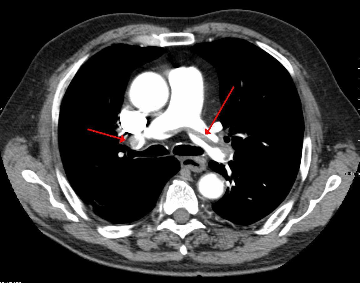
The stable patient still goes on for the scan that shows the clot. A CT of the lung arteries lights up the clot directly and confirms what the strained right ventricle suggested. A clinician reads the bedside study as the first step and the CT as the one that settles it. The echo raised the suspicion and sorted the risk. The CT then names the clot and its reach. A clinician sends the patient for the lung scan once the pressure is steady enough to travel. The two studies work in order, one fast at the bedside, the other firm down the hall. A clinician marks the time of each step, since the order of the echo and the scan shapes how fast the treatment can start. A blood thinner is the floor under every case, started the moment the clot is likely, held back only where bleeding forbids it.
The veins, the lungs, and the days after
The clot that strained the heart usually started in a deep vein of the leg. A clinician turns the probe to the big veins behind the knee and in the groin and presses down on each. The test is simple, whether the vein flattens under gentle pressure. A vein packed with clot holds its round shape and stays open under the probe. A clot found in the leg of a breathless patient backs the case for an embolism in the lung. A clinician reads the legs as the place the clot set out from. A clinician presses at two places, the groin and behind the knee, where the deep veins lie close enough to read. A vein that refuses to flatten at either point marks a clot a clinician can see without reaching the lung. A clinician reads a clot in the leg as the same disease the heart is showing, one process caught at both ends.
The lungs give their own answers under the probe. A clinician sweeps the chest for the signs that point away from a clot, since a collapsed lung, a wide pneumonia, or a lung waterlogged by heart failure can each cause the same breathlessness. A clear lung field keeps the focus on the right heart and the clot. A small wedge of dead lung at the surface, where an embolism has cut off the blood, sometimes shows as a shallow defect against the chest wall. A clinician folds the lung read into the same focused study. The chest sweep widens the search past the heart to the other causes of a sudden struggle to breathe. A clinician reads thin white lines rising from the lung surface as a sign of fluid in the lung, the mark of heart failure behind the breathlessness. A small pocket of fluid at the lung base can appear alongside the clot itself. A clinician reads the lung and the heart in one sweep, since the same probe answers both in the breathless patient.
The right heart recovers over the days after the clot is treated. A clinician brings the probe back to the bedside to watch the swollen right ventricle settle toward its normal size. A blood thinner holds new clot off and gives the body time to break the old one down. The pressure on the right side falls week by week. A clinician reads a shrinking right ventricle and a falling lung pressure as the sign the treatment is working. The septum rounds back out once the load comes off the right side. A clinician times these follow-up scans to the treatment and reads each against the first. A clinician sets a follow-up at a few weeks, the window where a recovering right heart has had time to settle. A clinician reads the recovery as a matter of days to weeks, the pace at which the freed lung lets the right heart unload.
A right ventricle that stays strained weeks later carries a warning. A clinician who finds a wide right ventricle and a high lung pressure long after the clot raises the question of a lasting blockage. Some clots never fully clear. The lung arteries stay narrowed for good and hold the pressure high. A clinician reads a strain that does not settle as a reason to look for chronic disease in the lung vessels. The follow-up scan, read against the first, shows whether the heart has recovered. A clinician sends the patient on for the deeper work-up when the right heart stays loaded. A clinician reads a lung pressure that stays high past three months as the line where chronic disease takes over from a single clot. A heart still loaded that late has earned a closer look at the lung vessels.
A pulmonary embolism hides its clot in the lung, far from any probe on the chest. A handheld reads the mark the clot leaves on the right heart, the strain, the dilation, the flattened septum, and the raised pressure. A clinician uses that read to judge the danger, to guide the crashing patient’s treatment, and to follow the heart back to health. The CT names the clot once the patient can travel for it. A clinician reads the right heart as the place the embolism shows its hand, and acts on what it shows. A clinician hands on a clear account of the right heart, its size, its pressure, and its trend, for whoever reads the patient next. A clinician writes the right-heart numbers in plain terms, so the next reader picks up the thread without a second guess.
Common questions about pulmonary embolism on a handheld scan
How does a pulmonary embolism show on a handheld scan?
It shows through the strain the clot throws onto the right side of the heart. A clinician reads a right ventricle that has swelled to match or outgrow the left, a septum flattened into a D-shape, a still free wall, and a raised pressure in the lung arteries. These signs together point to an acute load on the right heart. The scan reads this strain, since the clot itself sits out of reach in the lung.
Can the scan see the clot itself?
Rarely. The clot lodges in the arteries of the lung, beyond the reach of a probe on the chest. A clinician reads its effect on the right ventricle instead. Now and then a clot caught in transit shows as a worm-like shape moving through the right atrium or ventricle, a rare find that signals real danger. The clot in the lung is named on a CT of the lung arteries.
What is McConnell’s sign?
It is a pattern of motion in the strained right ventricle. The free wall falls still along much of its length. The patch near the apex keeps its normal squeeze, the one part the strain spares. McConnell first described this split as a pointer to an acute clot in the lung. A clinician reads it as a strong hint, weighed with the dilated chamber and the rest of the picture.
Does a normal scan rule out a pulmonary embolism?
No. A small clot can settle in the lung and never strain the right heart, so a normal right ventricle does not clear a suspicious patient. A clinician treats a normal scan as one piece of the picture and carries the work-up on. The CT of the lung arteries settles the case where the suspicion holds.
How does the scan change treatment?
It sorts the danger and guides the urgent steps. A strained right ventricle in a stable patient lifts the risk band and calls for a closer watch. In a crashing patient, a strained right ventricle supports a clot-busting drug on the spot, before any trip to the scanner. A clinician reads the scan to act fast where the patient cannot wait.

About the Author
Julien Mercier
Senior R&D Engineer · Medical Ultrasound Transducer Development
Senior R&D Engineer with an M.S. in Applied Physics and over 15 years of experience in medical ultrasound transducer development, specializing in the design verification and performance testing of high-frequency imaging transducers. Currently leading the development and verification of the company’s next-generation high-frequency linear-array transducer, responsible for imaging performance evaluation and reliability analysis in preclinical testing. Brings extensive hands-on experience in piezoelectric element tuning, beamforming parameter optimization, and system-level performance testing.